

Vol 44 # 2 June 2012 - Kma.org.kw
Vol 44 # 2 June 2012 - Kma.org.kw
Vol 44 # 2 June 2012 - Kma.org.kw

You also want an ePaper? Increase the reach of your titles
YUMPU automatically turns print PDFs into web optimized ePapers that Google loves.
151<br />
Bilateral Massive Angiomyolipomatosis Associated with Tuberous Sclerosis<br />
<strong>June</strong> <strong>2012</strong><br />
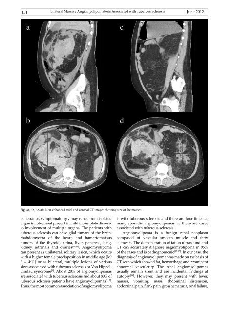
Fig. 3a, 3b, 3c, 3d: Non-enhanced axial and coronal CT images showing size of the masses<br />
penetrance, symptomatology may range from isolated<br />
<strong>org</strong>an involvement present in mild incomplete disease,<br />
to involvement of multiple <strong>org</strong>ans. The patients with<br />
tuberous sclerosis can have glial tumors of the brain,<br />
rhabdomyoma of the heart, and hamartomatous<br />
tumors of the thyroid, retina, liver, pancreas, lung,<br />
kidney, adrenals and ovaries [10,11] . Angiomyolipoma<br />
can present as unilateral, solitary lesion, which occurs<br />
with a higher female predisposition in middle age (M:<br />
F = 4:11) or as bilateral, multiple lesions of various<br />
sizes associated with tuberous sclerosis or Von Hippel-<br />
Lindau syndrome [1] . About 20% of angiomyolipomas<br />
are associated with tuberous sclerosis and about 80% of<br />
tuberous sclerosis patients have angiomyolipomas [2, 3] .<br />
Thus, the most common association of angiomyolipoma<br />
is with tuberous sclerosis and there are four times as<br />
many sporadic angiomyolipomas as there are cases<br />
associated with tuberous sclerosis.<br />
Angiomyolipoma is a benign renal neoplasm<br />
composed of vascular smooth muscle and fatty<br />
elements. The demonstration of fat on ultrasound and<br />
CT can accurately diagnose angiomyolipoma in 95%<br />
of the cases and is pathognomonic [12,13] . In our case, the<br />
diagnosis of angiomyolipoma was made on the basis of<br />
CT scan which showed fat, hemorrhage and prominent<br />
abnormal vascularity. The renal angiomyolipomas<br />
usually remain silent and are incidental findings at<br />
autopsy [14] . However, they may present with fever,<br />
nausea, vomiting, mass, abdominal distension,<br />
abdominal pain, flankpain, gross hematuria, renal failure,


