

Vol 43 # 2 June 2011 - Kma.org.kw
Vol 43 # 2 June 2011 - Kma.org.kw
Vol 43 # 2 June 2011 - Kma.org.kw

Create successful ePaper yourself
Turn your PDF publications into a flip-book with our unique Google optimized e-Paper software.
122<br />
Pattern of Chromosomal Abnormalities in Pediatric Acute Lymphoblastic Leukemia (ALL)<br />
<strong>June</strong> <strong>2011</strong><br />
Fig. 3: G-Banded male karyotype in ALL patient showing multiple<br />
whole chromosomal deletions such as deletion of Chr 2, 3, 4, 12, 13,<br />
15, 16, 17&Y (arrow). This is an example of numerical chromosomal<br />
abnormality in ALL [37, X, hypodiploidy and aneuploidy] (G-T-G<br />
Banding, 40X).<br />
cytogenetic techniques such as fluorescence in-situ<br />
hybridization (FISH). The FISH technique have become<br />
an integral part of cytogenetic analysis and must be<br />
regarded as complementary, not replacement tools [27,<br />
28]<br />
.<br />
The t (12; 21) (p13, q22) is the most common<br />
structural abnormality among pediatric B - ALL and<br />
carries a favorable prognosis. However, this can be<br />
missed by routine low resolution G-banding technique<br />
and hence remain cryptic [8] . In a recent study, by using<br />
conventional cytogenetics and FISH technique [28] , t (12;<br />
21) (q13; q 22) was found to be the most common (22%),<br />
followed in frequency, by 9p abnormalities (10%), t<br />
(1;19) (8%), t (9; 22) (8%), and 11q23 abnormality (5%).<br />
Rare translocations such as t (5;12), t (14,19), t (12;16),<br />
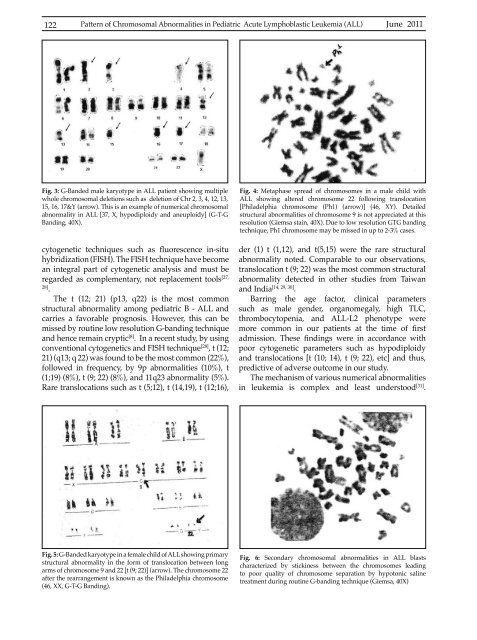
Fig. 4: Metaphase spread of chromosomes in a male child with<br />
ALL showing altered chromosome 22 following translocation<br />
[Philadelphia chromosome (Ph1) (arrow)] (46, XY). Detailed<br />
structural abnormalities of chromosome 9 is not appreciated at this<br />
resolution (Giemsa stain, 40X). Due to low resolution GTG banding<br />
technique, Ph1 chromosome may be missed in up to 2-3% cases.<br />
der (1) t (1,12), and t(5,15) were the rare structural<br />
abnormality noted. Comparable to our observations,<br />
translocation t (9; 22) was the most common structural<br />
abnormality detected in other studies from Taiwan<br />
and India [14, 29, 30] .<br />
Barring the age factor, clinical parameters<br />
such as male gender, <strong>org</strong>anomegaly, high TLC,<br />
thrombocytopenia, and ALL-L2 phenotype were<br />
more common in our patients at the time of first<br />
admission. These findings were in accordance with<br />
poor cytogenetic parameters such as hypodiploidy<br />
and translocations [t (10; 14), t (9; 22), etc] and thus,<br />
predictive of adverse outcome in our study.<br />
The mechanism of various numerical abnormalities<br />
in leukemia is complex and least understood [31] .<br />
Fig. 5: G-Banded karyotype in a female child of ALL showing primary<br />
structural abnormality in the form of translocation between long<br />
arms of chromosome 9 and 22 [t (9; 22)] (arrow). The chromosome 22<br />
after the rearrangement is known as the Philadelphia chromosome<br />
(46, XX, G-T-G Banding).<br />
Fig. 6: Secondary chromosomal abnormalities in ALL blasts<br />
characterized by stickiness between the chromosomes leading<br />
to poor quality of chromosome separation by hypotonic saline<br />
treatment during routine G-banding technique (Giemsa, 40X)


