

Vol 43 # 2 June 2011 - Kma.org.kw
Vol 43 # 2 June 2011 - Kma.org.kw
Vol 43 # 2 June 2011 - Kma.org.kw

You also want an ePaper? Increase the reach of your titles
YUMPU automatically turns print PDFs into web optimized ePapers that Google loves.
<strong>June</strong> <strong>2011</strong><br />
KUWAIT MEDICAL JOURNAL 131<br />
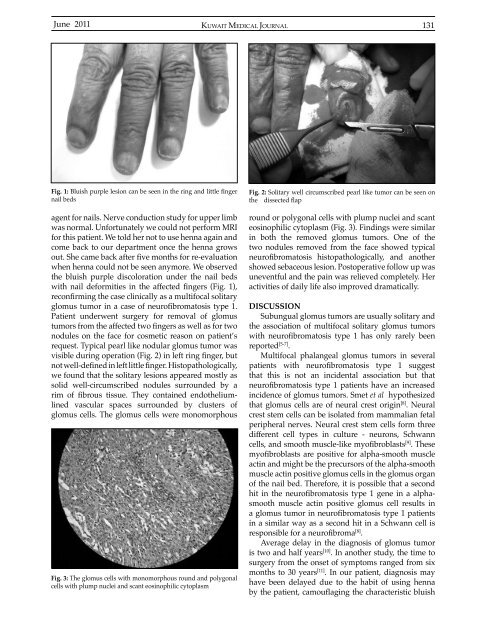
Fig. 1: Bluish purple lesion can be seen in the ring and little finger<br />
nail beds<br />
agent for nails. Nerve conduction study for upper limb<br />
was normal. Unfortunately we could not perform MRI<br />
for this patient. We told her not to use henna again and<br />
come back to our department once the henna grows<br />
out. She came back after five months for re-evaluation<br />
when henna could not be seen anymore. We observed<br />
the bluish purple discoloration under the nail beds<br />
with nail deformities in the affected fingers (Fig. 1),<br />
reconfirming the case clinically as a multifocal solitary<br />
glomus tumor in a case of neurofibromatosis type 1.<br />
Patient underwent surgery for removal of glomus<br />
tumors from the affected two fingers as well as for two<br />
nodules on the face for cosmetic reason on patient’s<br />
request. Typical pearl like nodular glomus tumor was<br />
visible during operation (Fig. 2) in left ring finger, but<br />
not well-defined in left little finger. Histopathologically,<br />
we found that the solitary lesions appeared mostly as<br />
solid well-circumscribed nodules surrounded by a<br />
rim of fibrous tissue. They contained endotheliumlined<br />
vascular spaces surrounded by clusters of<br />
glomus cells. The glomus cells were monomorphous<br />
Fig. 3: The glomus cells with monomorphous round and polygonal<br />
cells with plump nuclei and scant eosinophilic cytoplasm<br />
Fig. 2: Solitary well circumscribed pearl like tumor can be seen on<br />
the dissected flap<br />
round or polygonal cells with plump nuclei and scant<br />
eosinophilic cytoplasm (Fig. 3). Findings were similar<br />
in both the removed glomus tumors. One of the<br />
two nodules removed from the face showed typical<br />
neurofibromatosis histopathologically, and another<br />
showed sebaceous lesion. Postoperative follow up was<br />
uneventful and the pain was relieved completely. Her<br />
activities of daily life also improved dramatically.<br />
DISCUSSION<br />
Subungual glomus tumors are usually solitary and<br />
the association of multifocal solitary glomus tumors<br />
with neurofibromatosis type 1 has only rarely been<br />
reported [5-7] .<br />
Multifocal phalangeal glomus tumors in several<br />
patients with neurofibromatosis type 1 suggest<br />
that this is not an incidental association but that<br />
neurofibromatosis type 1 patients have an increased<br />
incidence of glomus tumors. Smet et al hypothesized<br />
that glomus cells are of neural crest origin [8] . Neural<br />
crest stem cells can be isolated from mammalian fetal<br />
peripheral nerves. Neural crest stem cells form three<br />
different cell types in culture - neurons, Schwann<br />
cells, and smooth muscle-like myofibroblasts [9] . These<br />
myofibroblasts are positive for alpha-smooth muscle<br />
actin and might be the precursors of the alpha-smooth<br />
muscle actin positive glomus cells in the glomus <strong>org</strong>an<br />
of the nail bed. Therefore, it is possible that a second<br />
hit in the neurofibromatosis type 1 gene in a alphasmooth<br />
muscle actin positive glomus cell results in<br />
a glomus tumor in neurofibromatosis type 1 patients<br />
in a similar way as a second hit in a Schwann cell is<br />
responsible for a neurofibroma [8] .<br />
Average delay in the diagnosis of glomus tumor<br />
is two and half years [10] . In another study, the time to<br />
surgery from the onset of symptoms ranged from six<br />
months to 30 years [11] . In our patient, diagnosis may<br />
have been delayed due to the habit of using henna<br />
by the patient, camouflaging the characteristic bluish


