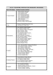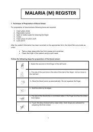
SOP â Malaria Microscopy - NVBDCP
SOP â Malaria Microscopy - NVBDCP
SOP â Malaria Microscopy - NVBDCP

You also want an ePaper? Increase the reach of your titles
YUMPU automatically turns print PDFs into web optimized ePapers that Google loves.
Quality Assurance of <strong>Malaria</strong> Diagnostic tests7.1.4 Bright field compound microscopeThe common microscope that is suited to see and study the microorganism routinely is thetypical compound microscope, either monocular or binocular. Here the microscopic field orarea observed is brightly lit and the objects under study appear darker. Generally, thesemicroscopes produce useful magnification of about upto 1000 times than the naked eye.• Monocular: monocular microscopes have single eye piece and are convenient foruse by beginners.• Binocular: binocular microscopes have two eye pieces. They are recommendedwhere much work has to be done, as this microscope causes less eye strain andfatigue.7.1.5 Parts of a microscope7.1.5.1. Mechanical7.1.5.1.1 StandIt forms the base of the microscope. It consists of a vertical pillar supported on a horse shoeshaped base or foot. It gives stability to the microscope. The stand is attached to the arm orlimb by the hinged (inclination) joint, which can be adjusted to any convenient angle. Thelimb or arm carries the illuminating apparatus, the stage and the observation tube. It alsoserves as a handle.7.1.5.1.2 StageIt is a platform with a circular aperture in the center. Stages are usually of two types:• a fixed stage in which the object is fixed by clips as in a monocular microscopy.• a mechanical stage in which the object can be moved to the desirable distance,either sideways or forward and backward, as in binocular microscope. This typeof stage is preferable for examination of a blood film or to locate a particular pointin the object.7.1.5.1.3 Focussing knobsThese are located on the side of the microscope; outermost is the course focus andinnermost is the fine focus. On the binocular microscopes, these knobs control up/downmovement of the stage.7.1.5. 2. Magnifying partsThese are eyepieces and objectives. They are kept separated in a graduated tube.7.1.5.2.1 Ocular lens or eyepieceUsually of 6x or 10x magnification. Only one eyepiece is present in a monocular microscopeand two in case of a binocular microscope.<strong>SOP</strong> – <strong>Malaria</strong> <strong>Microscopy</strong>© Copyright to Dte. <strong>NVBDCP</strong> Only. Any modification are prohibited