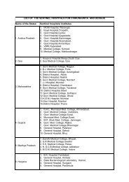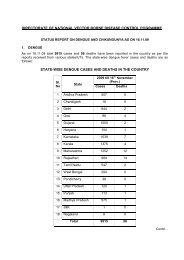
SOP â Malaria Microscopy - NVBDCP
SOP â Malaria Microscopy - NVBDCP
SOP â Malaria Microscopy - NVBDCP

You also want an ePaper? Increase the reach of your titles
YUMPU automatically turns print PDFs into web optimized ePapers that Google loves.
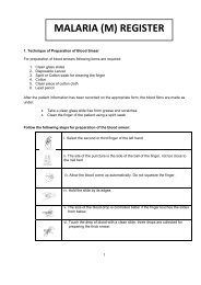
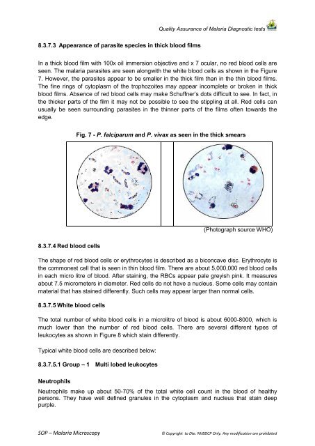
Quality Assurance of <strong>Malaria</strong> Diagnostic tests8.3.7.3 Appearance of parasite species in thick blood filmsIn a thick blood film with 100x oil immersion objective and x 7 ocular, no red blood cells areseen. The malaria parasites are seen alongwith the white blood cells as shown in the Figure7. However, the parasites appear to be smaller in the thick film than in the thin blood films.The fine rings of cytoplasm of the trophozoites may appear incomplete or broken in thickblood films. Absence of red blood cells may make Schuffner’s dots difficult to see. In fact, inthe thicker parts of the film it may not be possible to see the stippling at all. Red cells canusually be seen surrounding parasites in the thinner parts of the films often towards theedge.Fig. 7 - P. falciparum and P. vivax as seen in the thick smears(Photograph source WHO)8.3.7.4 Red blood cellsThe shape of red blood cells or erythrocytes is described as a biconcave disc. Erythrocyte isthe commonest cell that is seen in thin blood film. There are about 5,000,000 red blood cellsin each micro litre of blood. After staining, the RBCs appear pale greyish pink. It measuresabout 7.5 micrometers in diameter. Red cells do not have a nucleus. Some cells may containmaterial that has stained differently. Such cells may appear larger than normal cells.8.3.7.5 White blood cellsThe total number of white blood cells in a microlitre of blood is about 6000-8000, which ismuch lower than the number of red blood cells. There are several different types ofleukocytes as shown in Figure 8 which stain differently.Typical white blood cells are described below:8.3.7.5.1 Group – 1 Multi lobed leukocytesNeutrophilsNeutrophils make up about 50-70% of the total white cell count in the blood of healthypersons. They have well defined granules in the cytoplasm and nucleus that stain deeppurple.<strong>SOP</strong> – <strong>Malaria</strong> <strong>Microscopy</strong>© Copyright to Dte. <strong>NVBDCP</strong> Only. Any modification are prohibited