SOP â Malaria Microscopy - NVBDCP
SOP â Malaria Microscopy - NVBDCP
SOP â Malaria Microscopy - NVBDCP

You also want an ePaper? Increase the reach of your titles
YUMPU automatically turns print PDFs into web optimized ePapers that Google loves.
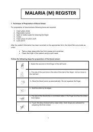
Quality Assurance of <strong>Malaria</strong> Diagnostic testsThe overall magnification achieved by the three objectives, 10x, 40x and 100x, when usedwith eye piece of 100x, 400 x and 1000 x, respectively.7.1.6.2. Resolution:Merely magnifying an object without a simultaneous increase in the amount of detail seenwill not provide the viewer with a good image. The ability of a microscope (or eye) to see thedetails is a function of its resolving power. Resolving power is defined as the minimumdistance between two objects at which the objects can just be distinguished as separate andis a function of the wavelength of light used and the quality of the optics. In general, theshorter the wavelength of the light source, the higher the resolution of the microscope.Working distance is the distance between the objective lens and the specimen. At lowmagnification, the working distance is relatively long. As the magnification is increased, theworking distance decreases dramatically. Oil immersion lenses practically touch thespecimen. Be aware of this change in working distance with increasing magnification so asto prevent damage to your specimens.7.1.7 Focussing procedure7.1.7.1 Low / high power focussing• turn on the light source.• switch to the 10x objective lens.• turn the coarse focus to raise the nose piece.• place the specimen slide on the stage and secure in the proper position. Look atthe slide and place it so that the specimen is over the light aperture in the stage.• lower the objective lens to lower limit (close to slide). Raise the lens using thecoarse focus knob until you see the image come into focus Adjust fine focussimilarly.• center the image and adjust the light using the diaphragm.• readjust diaphragm if needed.• now switch objectives to the 40x, if a higher magnification is needed. Readjustfine focus and light (diaphragm), as needed.The microscopes should be par focal which means that when you switch from low (100x)to high (400x) power, a focused image at low power will remain more or less in focus at thehigher power. Most likely the fine focus and diaphragm have to be readjusted slightly.7.1.7.2 Procedure for using oil immersion lens• locate the region of interest of the blood smear and center it with 40x objective.• then the objective lens is raised to its limit (i.e., maximize the distance betweenstage and objectives) and swing the lens out of the way, about half way to the nextposition.• place a small drop of immersion oil carefully, placed directly on the bloods smearover the center of the region of interest.• rotate the oil immersion objective into position carefully and while looking from theside, lower it using the coarse focus knob until the lens just makes contact with theoil drop. The drop leaps up into a column as the contact is made.• lower the lens a smidgen more and then using the fine focus and looking throughthe ocular lens, focus on the specimen.• when done, clean lens with lens paper until no more oil comes off and clean slide ifit is to be saved.<strong>SOP</strong> – <strong>Malaria</strong> <strong>Microscopy</strong>© Copyright to Dte. <strong>NVBDCP</strong> Only. Any modification are prohibited