

Color Atlas of Hematology - Practical Microscopic and Clinical ...
Color Atlas of Hematology - Practical Microscopic and Clinical ...
Color Atlas of Hematology - Practical Microscopic and Clinical ...
- No tags were found...

You also want an ePaper? Increase the reach of your titles
YUMPU automatically turns print PDFs into web optimized ePapers that Google loves.
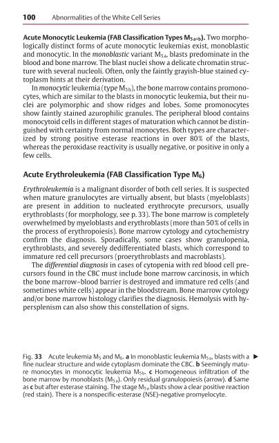
100Abnormalities <strong>of</strong> the White Cell SeriesAcute Monocytic Leukemia (FAB Classification Types M 5 a+b). Two morphologicallydistinct forms <strong>of</strong> acute monocytic leukemias exist, monoblastic<strong>and</strong> monocytic. In the monoblastic variant M 5a , blasts predominate in theblood <strong>and</strong> bone marrow. The blast nuclei show a delicate chromatin structurewith several nucleoli. Often, only the faintly grayish-blue stained cytoplasmhints at their derivation.In monocytic leukemia (type M 5b ), the bone marrow contains promonocytes,which are similar to the blasts in monocytic leukemia, but their nucleiare polymorphic <strong>and</strong> show ridges <strong>and</strong> lobes. Some promonocytesshow faintly stained azurophilic granules. The peripheral blood containsmonocytoid cells in different stages <strong>of</strong> maturation which cannot be distinguishedwith certainty from normal monocytes. Both types are characterizedby strong positive esterase reactions in over 80% <strong>of</strong> the blasts,whereas the peroxidase reactivity is usually negative, or positive in only afew cells.Acute Erythroleukemia (FAB Classification Type M 6 )Erythroleukemia is a malignant disorder <strong>of</strong> both cell series. It is suspectedwhen mature granulocytes are virtually absent, but blasts (myeloblasts)are present in addition to nucleated erythrocyte precursors, usuallyerythroblasts (for morphology, see p. 33). The bone marrow is completelyoverwhelmed by myeloblasts <strong>and</strong> erythroblasts (more than 50% <strong>of</strong> cells inthe process <strong>of</strong> erythropoiesis). Bone marrow cytology <strong>and</strong> cytochemistryconfirm the diagnosis. Sporadically, some cases show granulopenia,erythroblasts, <strong>and</strong> severely dedifferentiated blasts, which correspond toimmature red cell precursors (proerythroblasts <strong>and</strong> macroblasts).The differential diagnosis in cases <strong>of</strong> cytopenia with red blood cell precursorsfound in the CBC must include bone marrow carcinosis, in whichthe bone marrow–blood barrier is destroyed <strong>and</strong> immature red cells (<strong>and</strong>sometimes white cells) appear in the bloodstream. Bone marrow cytology<strong>and</strong>/or bone marrow histology clarifies the diagnosis. Hemolysis with hypersplenismcan also show this constellation <strong>of</strong> signs.Fig. 33 Acute leukemia M 5 <strong>and</strong> M 6. a In monoblastic leukemia M 5 a, blasts with a fine nuclear structure <strong>and</strong> wide cytoplasm dominate the CBC. b Seemingly maturemonocytes in monocytic leukemia M 5 b. c Homogeneous infiltration <strong>of</strong> thebone marrow by monoblasts (M 5 a). Only residual granulopoiesis (arrow). d Sameas c but after esterase staining. The stage M 5 a blasts show a clear positive reaction(red stain). There is a nonspecific-esterase (NSE)-negative promyelocyte.


