- Page 3 and 4: iiiColor Atlas of HematologyPractic
- Page 5 and 6: vPrefaceOur Current EditionAlthough
- Page 7 and 8: viiContentsPhysiology and Pathophys
- Page 9 and 10: ContentsixAcute Lymphoblastic Leuke
- Page 12 and 13: 2Physiology and Pathophysiology of
- Page 14 and 15: 4Physiology and Pathophysiology of
- Page 16 and 17: 6Physiology and Pathophysiology of
- Page 18 and 19: 8Physiology and Pathophysiology of
- Page 20 and 21: 10Physiology and Pathophysiology of
- Page 22 and 23: 12Physiology and Pathophysiology of
- Page 24 and 25: 14Physiology and Pathophysiology of
- Page 26 and 27: 16Physiology and Pathophysiology of
- Page 28 and 29: 18Physiology and Pathophysiology of
- Page 30 and 31: 20Physiology and Pathophysiology of
- Page 32 and 33: 22Physiology and Pathophysiology of
- Page 34 and 35: 24Physiology and Pathophysiology of
- Page 36 and 37: 26Physiology and Pathophysiology of
- Page 39 and 40: Normal Cells of the Blood andHemato
- Page 41: Normally erythropoiesis takes place
- Page 44 and 45: 34Normal Cells of the Blood and Hem
- Page 46 and 47: 36Normal Cells of the Blood and Hem
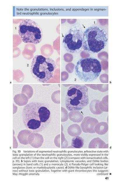
- Page 49: Advancing nuclear contraction and s
- Page 54 and 55: 44Normal Cells of the Blood and Hem
- Page 56 and 57: 46Normal Cells of the Blood and Hem
- Page 58 and 59: 48Normal Cells of the Blood and Hem
- Page 60 and 61: 50Normal Cells of the Blood and Hem
- Page 62 and 63: 52Normal Cells of the Blood and Hem
- Page 64 and 65: 54Normal Cells of the Blood and Hem
- Page 66 and 67: 56Normal Cells of the Blood and Hem
- Page 68 and 69: 58Normal Cells of the Blood and Hem
- Page 71 and 72: Abnormalities of the White Cell Ser
- Page 73 and 74: Predominance of Mononuclear Round t
- Page 75 and 76: Predominance of Mononuclear Round t
- Page 77 and 78: During lymphatic reactive states, v
- Page 79 and 80: Extreme transformation of lymphocyt
- Page 81 and 82: Predominance of Mononuclear Round t
- Page 83 and 84: Predominance of Mononuclear Round t
- Page 85 and 86: Monotonous proliferation of small l
- Page 87 and 88: Atypical lymphocytes are not part o
- Page 89 and 90: Deep nuclear indentation suggests f
- Page 91 and 92: Cytoplasmic processes the main feat
- Page 93 and 94: Plasmacytoma cannot be diagnosed wi
- Page 95 and 96: Atypias and differential diagnoses
- Page 97 and 98: Bone marrow diagnosis is indicated
- Page 99 and 100: Conspicuously large numbers of mono
- Page 101 and 102:
Predominance of Mononuclear Round t
- Page 103 and 104:
Predominance of Mononuclear Round t
- Page 105 and 106:
Predominance of Mononuclear Round t
- Page 107 and 108:
Fundamental characteristic of acute
- Page 109 and 110:
The diagnosis of acute leukemia is
- Page 111 and 112:
Acute leukemias may also derive fro
- Page 113 and 114:
New WHO classification: AML with dy
- Page 115 and 116:
The cells in acute lymphocytic leuk
- Page 117 and 118:
In unexplained anemia and/or leukoc
- Page 119 and 120:
The classification of myelodysplasi
- Page 121 and 122:
Prevalence of Polynuclear (Segmente
- Page 123 and 124:
Predominance of the granulocytic li
- Page 125 and 126:
Prevalence of Polynuclear (Segmente
- Page 127 and 128:
Left shift as far as myeloblasts, p
- Page 129 and 130:
Bone marrow analysis is not obligat
- Page 131 and 132:
In the course of chronic myeloid le
- Page 133 and 134:
Enlarged spleen and presence of imm
- Page 135 and 136:
Eosinophilia and basophilia are usu
- Page 137 and 138:
Erythrocyte and ThrombocyteAbnormal
- Page 139 and 140:
Hypochromic Anemias129Insufficient
- Page 141 and 142:
Hypochromic Anemias131BSGIron Ferri
- Page 143 and 144:
Small, hemoglobin-poor erythrocytes
- Page 145 and 146:
Hypochromic erythrocytes of very va
- Page 147 and 148:
Hypochromic Anemias137Hypochromic S
- Page 149 and 150:
Hypochromic anemia without iron def
- Page 151 and 152:
Consistently elevated “young” e
- Page 153 and 154:
Distribution pattern and shape of e
- Page 155 and 156:
Conspicuous erythrocyte morphology
- Page 157 and 158:
Unexplained decrease in cell counts
- Page 159 and 160:
Hypochromic Anemias149Differential
- Page 161 and 162:
Thrombocytopenia with leukocytosis
- Page 163 and 164:
Conspicuous large erythrocytes sugg
- Page 165 and 166:
In older patients, myelodysplastic
- Page 167 and 168:
Small inclusions are usually a sign
- Page 169 and 170:
PlasmodiumfalciparumPlasmodiumvivax
- Page 171 and 172:
Conspicuous erythrocyte inclusions
- Page 173 and 174:
Bone marrow analysis contributes to
- Page 175 and 176:
Thrombocytes: increases, reductions
- Page 177 and 178:
Thrombocytes: increases, reductions
- Page 179 and 180:
Variant forms of thrombocyte and me
- Page 181 and 182:
Thrombocyte proliferation with larg
- Page 183 and 184:
Cytology of Organ Biopsiesand Exuda
- Page 185 and 186:
Lymph Node Cytology175Anamnesis- su
- Page 187 and 188:
Reactive lymph node hyperplasia and
- Page 189 and 190:
Lymph Node Cytology179Contact witha
- Page 191 and 192:
Epithelioid cells dominate the lymp
- Page 193 and 194:
In cases of non-Hodgkin lymphoma an
- Page 195 and 196:
Accessible cysts (e.g., branchial c
- Page 197 and 198:
Tumor cells can be identified in pl
- Page 199 and 200:
Viral, bacterial, and malignant men
- Page 201 and 202:
191IndexAActinomycosis 179Addison d
- Page 203 and 204:
Index193Disseminated intravascularc
- Page 205 and 206:
Index195blast crisis 120-121bone ma
- Page 207 and 208:
Index197in hemolytic anemia 140orth