Applied Superconductivity - Walther Meißner Institut - Bayerische ...
Applied Superconductivity - Walther Meißner Institut - Bayerische ...
Applied Superconductivity - Walther Meißner Institut - Bayerische ...
- No tags were found...
Create successful ePaper yourself
Turn your PDF publications into a flip-book with our unique Google optimized e-Paper software.
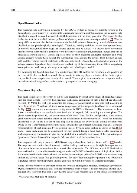
202 R. GROSS AND A. MARX Chapter 4Signal ReconstructionThe magnetic field distribution measured by the SQUID system is caused by currents flowing in thehuman body. Unfortunately, it is impossible to calculate the current distribution from the measured fielddistribution even if we could measure the field distribution with arbitrary precision. The reason for thatis the fact that the so-called inverse problem of electrodynamics has no unique solution. 106 That is,many different current distributions can create similar field patterns. Fortunately, not all of these currentdistributions are physiologically meaningful. Therefore, making additional model assumptions basedon medical background knowledge the inverse problem can be solved. All models have in commonthat the current distribution is assumed to be the sum of elementary physiological sources that can bemodeled as current dipoles. Current dipoles consist of short localized conductor segments and broadvolume currents flowing back through the surrounding tissue closing the circuit. Both the short currentpath and the volume current contribute to the magnetic field. Obviously, a detailed description of thevolume currents depends on the geometry and conductivity of the surrounding tissue. Often simplifyingassumptions are made as e.g. a homogeneous spherical or half-space model.By measuring the field distribution by a multichannel SQUID system the orientation and position ofthe current dipoles can be determined. For example, in this way the coordinates of the brain regionsresponsible for an epileptic attack can be determined. These regions in turn can be superimposed with athree-dimensional image of the brain obtained by magnetic resonance imaging.MagnetocardiographyThe heart signals are of the order of 100 pT and therefore by about three orders of magnitude largerthan the brain signals. However, fine structures with signal amplitudes of only a few pT are clinicallyrelevant. In MCG the goal is to determine the sources of pathological signals with high precision inthree dimensions. Therefore, all three vector components of the magnetic field have to be measured.In Fig. 4.29a a common measurement configuration in MCG is illustrated. The instantaneous heartaction is modeled by a current dipole associated with a magnetic field threading the sensor plane. Theplanar sensor loops detect B z , the z-component of the field. Thus, for this configuration, some sensorsyield positive and others negative values of the instantaneous field component B z . From the measureddistribution of B z values a so-called field map can be derived for every instant during the heart beat.Doing so one is interpolating between the values obtained in the different measuring channels. Commonrepresentations of such maps are by iso-contour lines or by false color scaling as shown in Fig. 4.29band c. Since such maps can be constructed for each instant during a heart beat, a video sequence ofsuch maps can be constructed to give the medical doctor a valuable impression of the spatio-temporaldynamics of the evolution of the magnetic field associated with the heart function.Two magnetic field map sequences (MFMS) covering a whole heart beat are shown in Fig. 4.29b and c.The sequence on the left is that of a volunteer with a healthy heart, whereas on the right the map sequenceof a patient is shown who suffered from ventricular tachycardia. The differences in field distributionsare considerable. It should be noted that a great variety of MFMS exists due to the biological variability.Each heart-healthy individual and each patient has its own finger print of an MFMS that may even varyin time and circumstances for a particular person. The art of interpreting these patterns is to identify thesignatures in these varying patterns that are clinically relevant indications of typical pathologies.While shielded rooms offer excellent conditions for basic biomagnetic research, unshielded operation ofSQUID systems is the ultimate goal from the commercial point of view. This is in particular true for MCGapplications. However, this goal is very hard to achieve and requires state-of-the-art noise suppression106 J. Sarvas, Phys. Med. Biol. 32, 11 (1987).© <strong>Walther</strong>-Meißner-<strong>Institut</strong>


