WIOMSA-CORDIO spawning book Full Doc 10 oct 13.pdf
WIOMSA-CORDIO spawning book Full Doc 10 oct 13.pdf
WIOMSA-CORDIO spawning book Full Doc 10 oct 13.pdf

Create successful ePaper yourself
Turn your PDF publications into a flip-book with our unique Google optimized e-Paper software.
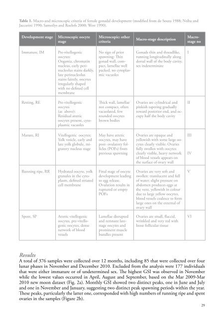
Table 1. Macro-and microscopic criteria of female gonadal development (modified from de Souza 1988; Ntiba andJaccarini 1990; Samoilys and Roelofs 2000; West 1990).Development stageMicroscopic oocytestageMicroscopic: othercriteriaMacro-stage descriptionMacrostagenoImmature, IMPre-vitellogenicoocytes:Oogonia, chromatinnucleus, early perinucleolusstains darkly,late perinucleolusstains faintly, oocytesirregularly shapedwith no defined cellmembraneNo sign of prior<strong>spawning</strong>: Thingonad wall, compact,lamellae wellpacked, no cytoplasmicvacuolesGonads thin and threadlike,running longitudinally alongdorsal wall of the body cavity,sex indeterminateIResting, REPre-vitellogenicoocytes(as above):Residual atreticoocytes present, cytoplasmicvacuolesThick wall, lamellaenot compact, oftenvacuolated, fewrounded oocytesbrown bodiesOvaries are cylindrical andpinkish tapering graduallytoward posterior end, and occupyhalf the body cavityIIMature, RIVitellogenic oocytes:Yolk vesicle, early andlate yolk globule, migratorynucleus stageMay have atreticoocytes, may havepost- ovulatory follicles(POFs) fromprevious <strong>spawning</strong>Ovaries are opaque andyellowish with some large oocytesclearly visible; Ovariesfully swollen with oocytesclearly visible, heavy networkof blood vessels appears onthe surface of ovary wallIIIIVRunning ripe, RRHydrated oocyte, yolkgranules in the cytoplasm,defined striatedcell membraneFinal stage of oocytedevelopment leadingto egg release.Ovulation results inruptured or emptyPOFsOvaries are very soft andswollen: translucent and fullof water; slight pressure onabdomen produces eggs atthe vent, yellowish in colourdue to large yellow oocytes,blood vessels coalesce to formlarge ones on the external ofovary wallVSpent, SPAtretic vitellogenicoocytes, pre-vitellogenicoocytes, densenetwork of bloodvesselsLamellae disruptedand remnant latestageoocytes andprominent musclebundles presentOvaries are small, flaccid,wrinkled and very red withloose follicular tissueVIResultsA total of 376 samples were collected over 12 months, including 85 that were collected over fourlunar phases in November and December 20<strong>10</strong>. Excluded from the analysis were 177 individualsthat were either immature or of undetermined sex. The highest GSI was observed in Novemberwhile the lowest values occurred in April, August and September, based on the Mar 2009-Mar20<strong>10</strong> new moon dataset (Fig. 2a). Monthly GSI showed two distinct peaks, one in June and Julyand one in November and January, suggesting two distinct peak <strong>spawning</strong> periods within the year.These peaks, particularly the latter one, corresponded with high numbers of running ripe and spentovaries in the samples (Figure 2b).29