Association
Magnetic Oxide Heterostructures: EuO on Cubic Oxides ... - JuSER
Magnetic Oxide Heterostructures: EuO on Cubic Oxides ... - JuSER
- No tags were found...
You also want an ePaper? Increase the reach of your titles
YUMPU automatically turns print PDFs into web optimized ePapers that Google loves.
3.2. In situ characterization techniques 41<br />
diffuse background, due to scattering from statistically distributed centers. Since the area<br />
on a sample surface in which constructive diffraction occurs is smaller than the coherence<br />
width of the electron beam, which amounts to ∼100 Å, a LEED probe is point-like. 79 Thus,<br />
LEED probes a microscopic area, while with RHEED one can “scan” the entire surface of the<br />
sample. In this work, LEED investigations are used to monitor the surface crystal structure<br />
by a qualitative comparison with simulated patterns of the LEEDpat software. 224 In practice,<br />
we use a three mesh VG Microtech RVL 640 Rear View LEED system.<br />
3.2.3. Auger electron spectroscopy (AES)<br />
Whenever functional heterostructures with the highest chemical quality of interfaces and<br />
surfaces are designed, an in situ analysis of the chemical composition and a quantification of<br />
surface oxidation or possible carbon contamination is essential. Among the non-destructive<br />
and element-selective chemical probing techniques, the Auger electron spectroscopy (AES) is<br />
well-established for analyzing the kinetic energy of secondary electrons. For light elements<br />
(Z 30), the Auger-Meitner cascade is the dominant emission process, when surface atoms<br />
are excited by X-rays or electrons. The external excitation leaves an electron core-hole (A),<br />
which is filled by an upper shell electron (B). This recombination energy is characteristic for<br />
the particular elemental atom and is used to emit an outer shell electron (C) carrying the<br />
characteristic kinetic energy<br />
E kin = E A − E B − E C − E vac .<br />
The specific Auger processes are named by the participating shells, e. g. KLM for transitions<br />
between the s and p orbitals and an emission from the d shell.<br />
<br />
<br />
<br />
<br />
<br />
<br />
<br />
<br />
<br />
<br />
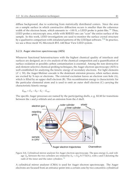
Figure 3.6.: Cylindrical mirror analyzer for Auger electron spectroscopy. The pass energy E 0 and voltage<br />
U p between the two cylinders are related by E 0 = eU p /0.77ln(b/a), with a and b denoting the<br />
radii of the inner and the outer cylinders. 79<br />
A cylindrical mirror analyzer (CMA) is used for Auger electron spectroscopy. The Auger<br />
electrons are focused from an entrance point into a certain cone by two concentric cylindrical


