Association
Magnetic Oxide Heterostructures: EuO on Cubic Oxides ... - JuSER
Magnetic Oxide Heterostructures: EuO on Cubic Oxides ... - JuSER
- No tags were found...

Create successful ePaper yourself
Turn your PDF publications into a flip-book with our unique Google optimized e-Paper software.
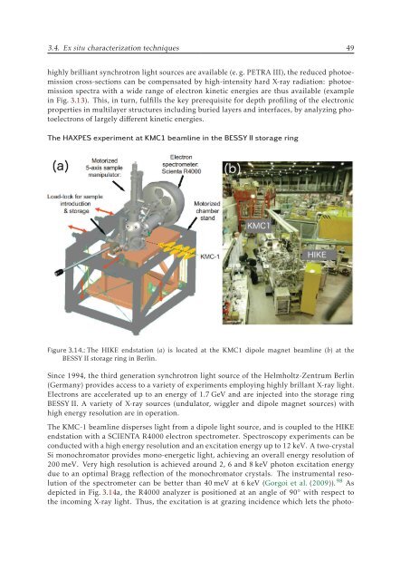
3.4. Ex situ characterization techniques 49<br />
highly brilliant synchrotron light sources are available (e. g. PETRA III), the reduced photoemission<br />
cross-sections can be compensated by high-intensity hard X-ray radiation: photoemission<br />
spectra with a wide range of electron kinetic energies are thus available (example<br />
in Fig. 3.13). This, in turn, fulfills the key prerequisite for depth profiling of the electronic<br />
properties in multilayer structures including buried layers and interfaces, by analyzing photoelectrons<br />
of largely different kinetic energies.<br />
The HAXPES experiment at KMC1 beamline in the BESSY II storage ring<br />
Figure 3.14.: The HIKE endstation (a) is located at the KMC1 dipole magnet beamline (b) at the<br />
BESSY II storage ring in Berlin.<br />
Since 1994, the third generation synchrotron light source of the Helmholtz-Zentrum Berlin<br />
(Germany) provides access to a variety of experiments employing highly brillant X-ray light.<br />
Electrons are accelerated up to an energy of 1.7 GeV and are injected into the storage ring<br />
BESSY II. A variety of X-ray sources (undulator, wiggler and dipole magnet sources) with<br />
high energy resolution are in operation.<br />
The KMC-1 beamline disperses light from a dipole light source, and is coupled to the HIKE<br />
endstation with a SCIENTA R4000 electron spectrometer. Spectroscopy experiments can be<br />
conducted with a high energy resolution and an excitation energy up to 12 keV. A two-crystal<br />
Si monochromator provides mono-energetic light, achieving an overall energy resolution of<br />
200 meV. Very high resolution is achieved around 2, 6 and 8 keV photon excitation energy<br />
due to an optimal Bragg reflection of the monochromator crystals. The instrumental resolution<br />
of the spectrometer can be better than 40 meV at 6 keV (Gorgoi et al. (2009)). 98 As<br />
depicted in Fig. 3.14a, the R4000 analyzer is positioned at an angle of 90 ◦ with respect to<br />
the incoming X-ray light. Thus, the excitation is at grazing incidence which lets the photo-