Undergrad_Book_16-18_Pge_View_Print_no print marks_compressed

You also want an ePaper? Increase the reach of your titles
YUMPU automatically turns print PDFs into web optimized ePapers that Google loves.
<strong>Undergrad</strong>uate Research at UMass Dartmouth<br />
107<br />
From previous research it has been found that<br />
patients with type 2 diabetes mellitus have an<br />
increased risk of bone fracture compared to <strong>no</strong>n-diabetics<br />
[1]. These patients have <strong>no</strong>rmal or high bone<br />
mass, which is typically beneficial for bone. This<br />
suggests factors other than bone mass, such as<br />
changes in bone quality, may play an important part<br />
in diabetic fractures. In this study, I looked at a possible<br />
method to inhibit harmful protein crosslinks<br />
that can accumulate in diabetic patients. I chose<br />
Vitamin B6 as the inhibitor because it showed<br />
promising results in rat bone [2]. However, it has<br />
never been tested in human bone. With this fact in<br />
mind, the goal of this project was to look at changes<br />
in protein crosslinks and mechanical properties of<br />
bone specimens after being placed in a simulated<br />
type 2 diabetic environment and to test how Vitamin<br />
B6 might prevent these changes.<br />
In the first part of this project, my main task was to<br />
work with human do<strong>no</strong>r bone (tibias or “shin bones”)<br />
that I cut and polished down to small testable sizes. I<br />
used a low-speed diamond blade saw and polishing<br />
machine to accomplish this task. Once all the specimens<br />
were ready, we then incubated them in control<br />
and type 2 diabetic environments (a chemical solution<br />
with ribose sugar) with and without Vitamin B6.<br />
The bone samples were incubated in these solutions<br />
for 10 days at 37ºC with pH maintained between 7.2<br />
and 7.6 to represent the human body environment.<br />
The second part of this project involved gathering<br />
data on the incubated bone specimens. When first<br />
looking at the post incubation samples, we saw a<br />
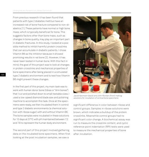
Jacob Aaronson (back) and John Riordan (front) making<br />
solutions for incubation of cortical bone specimens<br />
significant difference in color between ribose and<br />
control groups. Samples in ribose solutions were<br />
brown, which indicates a buildup of the protein<br />
crosslinks. Meanwhile control groups had <strong>no</strong><br />
significant color change. A biochemical assay was<br />
run to measure the crosslink content, and cyclic<br />
reference point indentation (RPI) tests were used<br />
to measure the mechanical properties of bone<br />
after incubation.