ischaemic preconditioning of the human heart. - Leicester Research ...
ischaemic preconditioning of the human heart. - Leicester Research ...
ischaemic preconditioning of the human heart. - Leicester Research ...

You also want an ePaper? Increase the reach of your titles
YUMPU automatically turns print PDFs into web optimized ePapers that Google loves.
712 0" et &L<br />
h wcomMe geg in Obwa»d Hý OSwardkm<br />
AMweviafiom and Acrosqmw<br />
CK = creatine, Idnase<br />
DCD = diet-controlled diabetes<br />
IDD = insulin-dependent diabetes<br />
K,<br />
Tp = AT? -dependent potassium channels<br />
LVEF = left ventricular ejection firaction<br />
MIT = 3-[4,5-dim&thyIthiazol-2-yI1-2,5<br />
diphenyltetrazolium. bromide<br />
NIDD = noninsulin-dependent diabetes<br />
PC = <strong>preconditioning</strong><br />
PKC =protein kinase C<br />
earlier shown, also in <strong>the</strong> rat <strong>heart</strong>, that myocardial infarc-<br />
tion is reduced in diabetes and that PC fin-<strong>the</strong>r increases <strong>the</strong><br />
protection <strong>of</strong> <strong>the</strong>se <strong>heart</strong>s. Ilere are very few studies in<br />
<strong>human</strong> diabetic tissue. Cleveland et al. (16) used a func-<br />
tional isolated atrial ft-abeculae model and showed that PC<br />
did not confer any protection <strong>of</strong> <strong>the</strong> myocardium on patients<br />
taking long-term oral hypoglycemic agents. They hypo<strong>the</strong>sized<br />
that long-term inhibition <strong>of</strong> KA-rp channels with <strong>the</strong>se<br />
agents may be responsible for <strong>the</strong> excess cardiovascular<br />
mortality associated with diabetes.<br />
Heart failure is common in all forms <strong>of</strong> <strong>heart</strong> disease.<br />
Mechanical dysfunction <strong>of</strong> <strong>the</strong> failing <strong>heart</strong> is due to many<br />
factors, including neurohormonal disturbance, accumulation<br />
<strong>of</strong> extr; icellular matrix, alteration <strong>of</strong> excitation-contraction<br />
coupling and maladaptation <strong>of</strong> myocardial energetics (17).<br />
Very few studies have investigated <strong>the</strong> effects <strong>of</strong> <strong>the</strong> PC<br />
response in <strong>the</strong> failing myocardiurn in light <strong>of</strong> alterations in<br />
<strong>the</strong> cellular metabolic and biochemical pathways associated<br />
with <strong>heart</strong> failure. Cleveland et al. (3) showed in isolated<br />
ventricular tr-Abeculae from patients requiring <strong>heart</strong> trans-<br />
plantation that PC conferred protection. However, more<br />
recently Dekker et al. (18) have studied perfused papillary<br />
muscles from rabbits in which cardiac failure has been<br />
induced by a combination <strong>of</strong> pressure and volume overload-<br />
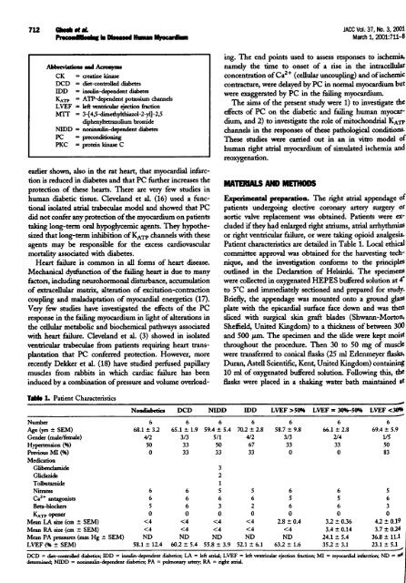
TaMe L Patient Characteristics<br />
JACC Vol. 37, No. 3,2001<br />
March 1,2001: 711-8<br />
ing. The end points used to assess responses to ischemia<br />
namely <strong>the</strong> time to onset <strong>of</strong> a rise in <strong>the</strong> intracellula<br />
concentration <strong>of</strong> Ca" (cellular uncoupling) and <strong>of</strong> ischerni<br />
contracture, were delayed by PC in normal myocardiurn but<br />
were exaggerated by PC in <strong>the</strong> fiding myocardium.<br />
The aims <strong>of</strong> <strong>the</strong> present study were 1) to investigate <strong>the</strong><br />
effiects <strong>of</strong> PC on <strong>the</strong> diabetic and failing <strong>human</strong> myocar<br />
dium, and 2) to investigate <strong>the</strong> role <strong>of</strong> mitochondrial. KAjT<br />
channels in <strong>the</strong> responses <strong>of</strong> <strong>the</strong>se pathological conditions<br />
These studies were carried out in an in vitro model <strong>of</strong><br />
<strong>human</strong> right atrial myocardium <strong>of</strong> simulated ischemia and<br />
reoxygenation.<br />
MATERUNLS MD METHODS<br />
preparation. The right atrial appendage <strong>of</strong><br />
patients undergoing elective coronary artery surgery or<br />
aortic valve replacement was obtained. Patients were ex-<br />
cluded if <strong>the</strong>y hýd enlarged right atriums, atrial arrhythmia<br />
or right ventricular failure, or were taking opioid analgesia<br />
Patient characteristics are detailed in Table 1. Local ethica<br />
committee approval was obtained for <strong>the</strong> harvesting tech-<br />
nique, and <strong>the</strong> investigation conforms to <strong>the</strong> principles<br />
outlined in <strong>the</strong> Declaration <strong>of</strong> Helsinki. The specimen<br />
were collected in oxygenated HEPES buffered solution at 4'<br />
to 5*C and immediately sectioned and prepared for study<br />
Briefly, <strong>the</strong> appendage was mounted onto a ground glass<br />
plate with <strong>the</strong> epicardial surface face down and was <strong>the</strong>n<br />
sliced with surgical skin graft blades (Shwann-Morton<br />
Sheffield, United Kingdom) to a thickness <strong>of</strong> between 300<br />
and 500 jAm. 'Me specimen and <strong>the</strong> slide were kept moist<br />
throughout <strong>the</strong> procedure. Then 30 to 50 mg <strong>of</strong> muscle<br />
were transferred to conical flasks (25 ml ErIenmeyer flasks<br />
Duran, Astell Scientific, Kent, United Kingdom) containing<br />
10 ml <strong>of</strong> oxygenated buffered solution. Following this, <strong>the</strong><br />
flasks were placed in a shaking water bath maintained at<br />
Noudiabetics DCD NIDD IDD LVEF >50% LVEF = 30%-50% LVEF