ARTIGO ORIGINAL99Dynamic ultra high speed Scheimpflug imagingfor assessing corneal biomechanical propertiesAvaliação Dinâmica com fotografia <strong>de</strong> Scheimpflug <strong>de</strong> altaveloci<strong>da</strong><strong>de</strong> para avaliar as proprie<strong>da</strong><strong>de</strong>s biomecânicas <strong>da</strong> córneaRenato Ambrósio Jr 1,2,3 , Isaac Ramos 1,2 , Allan Luz 2,3 , Fernando Correa Faria 1,2 , Andreas Steinmueller 4 ,Matthias Krug 4 , Michael W. Belin 5 , Cynthia Jane Roberts 6ABSTRACTObjective: To <strong>de</strong>scribe a novel technique for clinical characterization of corneal biomechanics using non-invasive dynamic imaging.Methods: Corneal <strong>de</strong>formation response during non contact tonometry (NCT) is monitored by ultra-high-speed (UHS) photography.The Oculus Corvis ST (Scheimpflug Technology; Wetzlar, Germany) has a UHS Scheimpflug camera, taking over 4,300 frames persecond and of a single 8mm horizontal slit, for monitoring corneal <strong>de</strong>formation response to NCT. The metered collimated air pulse orpuff has a symmetrical configuration and fixed maximal internal pump pressure of 25 kPa. The bidirectional mov<strong>em</strong>ent of the cornea inresponse to the air puff is monitored. Results: Measur<strong>em</strong>ent time is 30ms, with 140 frames acquired. Advanced algorithms for edge<strong>de</strong>tection of the front and back corneal contours are applied for every frame. IOP is calculated based on the first applanation moment.Deformation amplitu<strong>de</strong> (DA) is <strong>de</strong>termined as the highest displac<strong>em</strong>ent of the apex in the highest concavity (HC) moment. Applanationlength (AL) and corneal velocity (CVel) are recor<strong>de</strong>d during ingoing and outgoing phases. Conclusion: Corneal <strong>de</strong>formation can b<strong>em</strong>onitored during non contact tonometry. The parameters generated provi<strong>de</strong> clinical in vivo characterization of corneal biomechanicalproperties in two dimensions, which is relevant for different applications in Ophthalmology.Keywords: Biomechanics; Cornea/physiology; Corneal topography/methods; Tonometry, ocular/methodsRESUMOObjetivo: Descrever uma nova técnica para caracterização clínica <strong>da</strong>s proprie<strong>da</strong><strong>de</strong>s biomecânicas <strong>da</strong> córnea, usando sist<strong>em</strong>a <strong>de</strong>imag<strong>em</strong> dinâmica não invasivo. Métodos: A resposta <strong>de</strong> <strong>de</strong>formação <strong>da</strong> córnea é monitora<strong>da</strong> durante tonometria <strong>de</strong> não contatocom fotografia <strong>de</strong> Scheimpflug <strong>de</strong> altíssima veloci<strong>da</strong><strong>de</strong>. O Oculus Corvis ST (Scheimpflug Technology; Wetzlar, Germany) apresentauma câmera UHS Scheimpflug que adquire mais que 4.300 fotos por segundo com cobertura <strong>de</strong> 8mm horizontais para monitorar aresposta <strong>de</strong> <strong>de</strong>formação durante a tonometria <strong>de</strong> não contato por sopro <strong>de</strong> ar. O pulso <strong>de</strong> ar é muito b<strong>em</strong> controlado, apresentandouma configuração simétrica <strong>em</strong> sua pressão, com máxima pressão <strong>da</strong> bomba fixa <strong>de</strong> 25 kPa. O movimento bidirecional <strong>da</strong> córnea <strong>em</strong>resposta ao jato <strong>de</strong> ar é monitorado. Resultados: A medi<strong>da</strong> dura 30ms, com 140 fotos adquiri<strong>da</strong>s. Algorítmos avançados i<strong>de</strong>ntificamos limites anterior e posterior <strong>da</strong> córnea <strong>em</strong> ca<strong>da</strong> imag<strong>em</strong>. A pressão intraocular (PIO) é calcula<strong>da</strong> com base no primeiro momento<strong>de</strong> aplanação. A amplitu<strong>de</strong> <strong>de</strong> <strong>de</strong>formação é <strong>de</strong>termina<strong>da</strong> pelo maior <strong>de</strong>slocamento do ápice, durante o momento <strong>de</strong> maior concavi<strong>da</strong><strong>de</strong>.A extensão <strong>da</strong> aplanação e veloci<strong>da</strong><strong>de</strong> <strong>da</strong> córnea são medidos nas fases <strong>de</strong> entra<strong>da</strong> e saí<strong>da</strong>. Conclusão: A <strong>de</strong>formação <strong>da</strong> córneadurante a tonometria <strong>de</strong> sopro por pulso <strong>de</strong> ar po<strong>de</strong> ser monitora<strong>da</strong> <strong>em</strong> <strong>de</strong>talhe com sist<strong>em</strong>a <strong>de</strong> fotografia <strong>de</strong> altíssima veloci<strong>da</strong><strong>de</strong>.Os parâmetros gerados possibilitam caracterização clínica <strong>da</strong>s proprie<strong>da</strong><strong>de</strong>s biomecâmicas <strong>da</strong> córnea <strong>em</strong> duas dimensões. Taisachados têm relavância para diversas áreas <strong>da</strong> Oftalmologia.Descritores: Biomecânica; Córnea/fisiologia; Topografia <strong>da</strong> córnea/métodos; Tanometria ocular/métodos1Instituto <strong>de</strong> Olhos Renato Ambrósio – Rio <strong>de</strong> Janeiro (RJ), Brazil;2Rio <strong>de</strong> Janeiro Corneal Tomography and Biomechanics Study Group – Rio <strong>de</strong> Janeiro (RJ), Brazil;3Department of Ophthalmology, il;4Oculus GmbH - Wetzlar, Germany;5Ophthalmology, University of Arizona – Tucson, AZ, USA;6Ophthalmology, Department of Biomedical Engineering, The Ohio State University – Columbus, OH, USA.Financial Disclosure(s): Mr. Steinmueller, Mr. Krug are <strong>em</strong>ployee of Oculus; Dr. Ambrósio, Dr. Belin and Dr. Roberts are consultants for OculusRecebido para publicação <strong>em</strong>: 11/4/2012 - Aceito para publicação <strong>em</strong>: 3/12/2012Rev Bras Oftalmol. 2013; 72 (2): 99-102
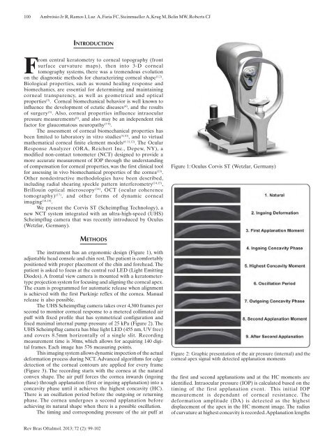
100Ambrósio Jr R, Ramos I, Luz A, Faria FC, Steinmueller A, Krug M, Belin MW, Roberts CJINTRODUCTIONFrom central keratometry to corneal topography (frontsurface curvature maps), then into 3-D cornealtomography syst<strong>em</strong>s, there was a tr<strong>em</strong>endous evolutionon the diagnostic methods for characterizing corneal shape (1,2) .Biological properties, such as wound healing response andbiomechanics, are essential for <strong>de</strong>termining and maintainingcorneal transparency, as well as geometrical and opticalproperties (3) . Corneal biomechanical behavior is well known toinfluence the <strong>de</strong>velopment of ectatic diseases (4) , and the resultsof surgery (5) . Also, corneal properties influence intraocularpressure measur<strong>em</strong>ents (6) , and also may be an in<strong>de</strong>pen<strong>de</strong>nt riskfactor for glaucomatous neuropathy (7-9) .The assessment of corneal biomechanical properties hasbeen limited to laboratory in vitro studies (4,10) , and to virtualmath<strong>em</strong>atical corneal finite el<strong>em</strong>ent mo<strong>de</strong>ls (6 11,12) . The OcularResponse Analyzer (ORA, Reichert Inc., Depew, NY), amodified non-contact tonometer (NCT) <strong>de</strong>signed to provi<strong>de</strong> amore accurate measur<strong>em</strong>ent of IOP through the un<strong>de</strong>rstandingof compensation for corneal properties, was the first clinical toolfor assessing in vivo biomechanical properties of the cornea (13) .Other non<strong>de</strong>structive methodologies have been <strong>de</strong>scribed,including radial shearing speckle pattern interferometry (14,15) ,Brillouin optical microscopy (16) , OCT (ocular coherencetomography) (17) , and other forms of dynamic cornealimaging (18,19) .We present the Corvis ST (Scheimpflug Technology), anew NCT syst<strong>em</strong> integrated with an ultra-high-speed (UHS)Scheimpflug camera that was recently introduced by Oculus(Wetzlar, Germany).Figure 1: Oculus Corvis ST (Wetzlar, Germany)METHODSThe instrument has an ergonomic <strong>de</strong>sign (Figure 1), withadjustable head console and chin rest. The patient is comfortablypositioned with proper plac<strong>em</strong>ent of the chin and forehead. Thepatient is asked to focus at the central red LED (Light EmittingDio<strong>de</strong>s). A frontal view camera is mounted with a keratometertypeprojection syst<strong>em</strong> for focusing and aligning the corneal apex.The exam is programmed for automatic release when alignmentis achieved with the first Purkinje reflex of the cornea. Manualrelease is also possible.The UHS Scheimpflug camera takes over 4,300 frames persecond to monitor corneal response to a metered collimated airpuff with fixed profile that has symmetrical configuration andfixed maximal internal pump pressure of 25 kPa (Figure 2). TheUHS Scheimpflug camera has blue light LED (455 nm, UV free)and covers 8.5mm horizontally of a single slit. Recordingmeasur<strong>em</strong>ent time is 30ms, which allows for acquiring 140 digitalframes. Each image has 576 measuring points.This imaging syst<strong>em</strong> allows dynamic inspection of the actual<strong>de</strong>formation process during NCT. Advanced algorithms for edge<strong>de</strong>tection of the corneal contours are applied for every frame(Figure 3). The recording starts with the cornea at the naturalconvex shape. The air puff forces the cornea inwards (ingoingphase) through applanation (first or ingoing applanation) into aconcavity phase until it achieves the highest concavity (HC).There is an oscillation period before the outgoing or returningphase. The cornea un<strong>de</strong>rgoes a second applanation beforeachieving its natural shape when there is a possible oscillation.The timing and corresponding pressure of the air puff atFigure 2: Graphic presentation of the air pressure (internal) and thecorneal apex signal with <strong>de</strong>tected applanation momentsthe first and second applanations and at the HC moments arei<strong>de</strong>ntified. Intraocular pressure (IOP) is calculated based on thetiming of the first applanation event. This initial IOPmeasur<strong>em</strong>ent is <strong>de</strong>pen<strong>da</strong>nt of corneal resistance. The<strong>de</strong>formation amplitu<strong>de</strong> (DA) is <strong>de</strong>tected as the highestdisplac<strong>em</strong>ent of the apex in the HC moment image. The radiusof curvature at highest concavity is recor<strong>de</strong>d. Applanation lengthsRev Bras Oftalmol. 2013; 72 (2): 99-102
- Page 1 and 2: Versão impressavol. 72 - nº 2 - M
- Page 3 and 4: 80Revista Brasileira de Oftalmologi
- Page 5 and 6: 82108 Efeitos de algumas drogas sob
- Page 7 and 8: 84Kara-Junior Ninúmeras vezes ante
- Page 9 and 10: 86Ambrósio Júnior Raplicação de
- Page 11 and 12: 88Monte FQ, Stadtherr NMINTRODUÇÃ
- Page 13 and 14: 90Monte FQ, Stadtherr NMFigura 1AFi
- Page 15 and 16: 92Monte FQ, Stadtherr NMtempo (3) .
- Page 17 and 18: 94Monte FQ, Stadtherr NM11. Höffli
- Page 19 and 20: 96Grandinetti AA, Dias J, Trautwein
- Page 21: 98Grandinetti AA, Dias J, Trautwein
- Page 25 and 26: 102Ambrósio Jr R, Ramos I, Luz A,
- Page 27 and 28: 104Caballero JCS, Centurion VINTROD
- Page 29 and 30: 106Caballero JCS, Centurion VDISCUS
- Page 31 and 32: ARTIGO ORIGINALEfeitos de algumas d
- Page 33 and 34: 110Almodin J, Almodin F, Almodin E,
- Page 35 and 36: 112 ARTIGO ORIGINALFluência do las
- Page 37 and 38: 114Lucena AR, Andrade NL, Lucena DR
- Page 39 and 40: 116RELATO DE CASOAsymptomatic ocula
- Page 41 and 42: 118Freitas LGA, Gabriel LAR, Isaac
- Page 43 and 44: 120Ticly FG, Lira RPC, Fulco EAM, V
- Page 45 and 46: 122RELATO DE CASOAssociation of mac
- Page 47 and 48: 124Tavares RLP, Novelli FJ, Nóbreg
- Page 49 and 50: 126Marback EF, Freitas MS, Spínola
- Page 51 and 52: 128RELATO DE CASONeurofibromatose t
- Page 53 and 54: 130Moraes FS, Santos WEM, Salomão
- Page 55 and 56: 132 ARTIGO DE REVISÃOAntifúngicos
- Page 57 and 58: 134Müller GG, Kara-José N, Castro
- Page 59 and 60: 136Müller GG, Kara-José N, Castro
- Page 61 and 62: 138Müller GG, Kara-José N, Castro
- Page 63 and 64: 140Müller GG, Kara-José N, Castro
- Page 65 and 66: 142 ARTIGO DE REVISÃOImportância
- Page 67 and 68: 144Rehder JRCL, Paulino LV, Paulino
- Page 69 and 70: 146Rehder JRCL, Paulino LV, Paulino
- Page 71 and 72: 148Instruções aos autoresA Revist
- Page 73:
150RevistaBrasileira deOftalmologia