RELATO DE CASO119Intravitreal ranibizumab as adjuvanttreatment for neovascular glaucomaRanibizumabe intravítreo como tratamentoadjuvante para glaucoma neovascularFlavia Gazze Ticly 1 , Rodrigo Pessoa Cavalcanti Lira 1 , Enzo Augusto Me<strong>de</strong>iros Fulco 1 , José Paulo Cabral <strong>de</strong>Vasconcelos 1ABSTRACTThe purpose of this study was to <strong>de</strong>scribe a prospective case series of 5 eyes treated with intravitreal ranibizumab injection for neovascularglaucoma (NVG). Five patients with clinically uncontrolled NVG secon<strong>da</strong>ry to proliferative diabetic retinopathy (4 patients) andcentral retinal vein occlusion (1 patient), non-responsive to maximal tolerable medication and panretinal photocoagulation, receivedintravitreal ranibizumab injection (0.5 mg). Patients were seen at 1st, 3rd and 7th <strong>da</strong>y after the ranibizumab injection and when it wasnecessary. Success was <strong>de</strong>fined as intraocular pressure (IOP) 21, <strong>de</strong>spite maximal tolerable medication, un<strong>de</strong>rwent trabeculectomy with 0.5mg/ml mitomycin C (MMC) for 1 minute. Failure was<strong>de</strong>fined as IOP > 21 mmHg, phthisis bulbi, loss of light perception or additional glaucoma surgery. The primary outcome was 6-monthIOP control. Mean IOP before the ranibizumab injection was 37 mmHg (7 mmHg SD). Two out of five eyes un<strong>de</strong>rwent only ranibizumabinjection, having an IOP control after the procedure. Three patients were submitted to trabeculectomy with MMC on the 7th <strong>da</strong>y afterthe injection. At 6-month follow-up, the mean IOP was 12mmHg (3 mmHg SD). All eyes showed regression of rubeosis iridis and IOPcontrol. Visual acuity improved in 2 eyes worsened in 1 eye, and r<strong>em</strong>ained stable in 2 eyes. These <strong>da</strong>ta suggest that intravitreal ranibizumabinjection may be a useful tool in the treatment of NVG.Keywords: Neovascular, glaucoma/drug therapy; Ch<strong>em</strong>otherapy, adjuvante; Intraocular pressure; Intravitreal injections; Antibodies,monoclonal/therapeutic use; Case reportsRESUMOO objetivo <strong>de</strong>ste estudo foi <strong>de</strong>screver uma série <strong>de</strong> casos prospectivos <strong>de</strong> 5 olhos tratados com ranibizumabe intravítreo paraglaucoma neovascular (GNV). Cinco pacientes com GNV refratário, secundário a retinopatia diabética proliferativa (4 pacientes)e oclusão <strong>de</strong> veia central <strong>da</strong> retina (1 paciente), não responsivos a terapia medicamentosa máxima tolera<strong>da</strong> e panfotocoagulação <strong>da</strong>retina, receberam ranibizumabe intravítreo (0,5 mg). Os pacientes foram vistos no 1º, 3º e 7º dia após a aplicação e conformenecessário. O sucesso foi <strong>de</strong>finido como pressão intraocular (PIO) d”21 mmHg, com ou s<strong>em</strong> uso <strong>de</strong> medicação antiglaucomatosa.Aqueles com PIO > 21 mmHg, apesar <strong>da</strong> medicação máxima tolera<strong>da</strong>, foram submetidos à trabeculectomia com mitomicina C(MMC) 0,5mg/mL por 1 minuto. Falência foi <strong>de</strong>fini<strong>da</strong> como PIO > 21 mmHg, phthisis bulbi, per<strong>da</strong> <strong>da</strong> percepção <strong>de</strong> luz ou necessi<strong>da</strong><strong>de</strong><strong>de</strong> cirurgia antiglaucomatosa adicional. O resultado primário avaliado foi o controle <strong>da</strong> PIO após 6 meses do procedimento. A PIOmédia antes <strong>da</strong> injeção era <strong>de</strong> 37 mmHg (DP=7 mmHg). Dois pacientes foram submetidos somente a injeção intravítrea <strong>de</strong>ranibizumabe, obtendo controle <strong>da</strong> PIO após o procedimento. Três pacientes foram submetidos à trabeculectomia com MMC no 7ºdia após a injeção. Após 6 meses <strong>de</strong> seguimento, a PIO média era <strong>de</strong> 12 mmHg (DP=3 mmHg). Todos os olhos mostraram regressão<strong>da</strong> rubeosis iriana e controle <strong>da</strong> PIO. A acui<strong>da</strong><strong>de</strong> visual melhorou <strong>em</strong> 2 olhos, piorou <strong>em</strong> 1 olho e permaneceu estável <strong>em</strong> 2 olhos.Estas informações suger<strong>em</strong> que a injeção intravítrea <strong>de</strong> ranibizumabe po<strong>de</strong> ser uma ferramenta útil no tratamento do GNV.Descritores: Glaucoma neovascular/quimioterapia; Quimioterapia adjuvante; Pressão intraocular; Injeções intravítreas;Anticorpos monoclonais/uso terapêutico; Relatos <strong>de</strong> casos1.Department of Ophthalmology, Universi<strong>da</strong><strong>de</strong> Estadual <strong>de</strong> Campinas (UNICAMP) – Campinas (SP), Brasil.Study carried out at <strong>de</strong>partment of Ophthalmology, Universi<strong>da</strong><strong>de</strong> Estadual <strong>de</strong> Campinas (UNICAMP) – Campinas (SP), Brasil.The authors <strong>de</strong>clare no conflicts of interestRecebido para publicação <strong>em</strong>: 5/10/2011 - Aceito para publicação <strong>em</strong>: 12/1/2012Rev Bras Oftalmol. 2013; 72 (2): 119-21
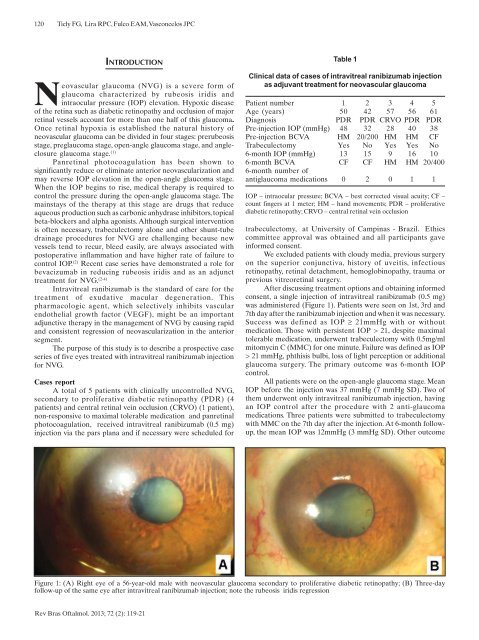
120Ticly FG, Lira RPC, Fulco EAM, Vasconcelos JPCINTRODUCTIONNeovascular glaucoma (NVG) is a severe form ofglaucoma characterized by rubeosis iridis andintraocular pressure (IOP) elevation. Hypoxic diseaseof the retina such as diabetic retinopathy and occlusion of majorretinal vessels account for more than one half of this glaucoma.Once retinal hypoxia is established the natural history ofneovascular glaucoma can be divi<strong>de</strong>d in four stages: prerubeosisstage, preglaucoma stage, open-angle glaucoma stage, and angleclosureglaucoma stage. (1)Panretinal photocoagulation has been shown tosignificantly reduce or eliminate anterior neovascularization andmay reverse IOP elevation in the open-angle glaucoma stage.When the IOP begins to rise, medical therapy is required tocontrol the pressure during the open-angle glaucoma stage. Th<strong>em</strong>ainstays of the therapy at this stage are drugs that reduceaqueous production such as carbonic anhydrase inhibitors, topicalbeta-blockers and alpha agonists. Although surgical interventionis often necessary, trabeculectomy alone and other shunt-tubedrainage procedures for NVG are challenging because newvessels tend to recur, bleed easily, are always associated withpostoperative inflammation and have higher rate of failure tocontrol IOP. (2) Recent case series have d<strong>em</strong>onstrated a role forbevacizumab in reducing rubeosis iridis and as an adjuncttreatment for NVG. (2-4)Intravitreal ranibizumab is the stan<strong>da</strong>rd of care for thetreatment of exu<strong>da</strong>tive macular <strong>de</strong>generation. Thispharmacologic agent, which selectively inhibits vascularendothelial growth factor (VEGF), might be an importantadjunctive therapy in the manag<strong>em</strong>ent of NVG by causing rapi<strong>da</strong>nd consistent regression of neovascularization in the anteriorsegment.The purpose of this study is to <strong>de</strong>scribe a prospective caseseries of five eyes treated with intravitreal ranibizumab injectionfor NVG.Cases reportA total of 5 patients with clinically uncontrolled NVG,secon<strong>da</strong>ry to proliferative diabetic retinopathy (PDR) (4patients) and central retinal vein occlusion (CRVO) (1 patient),non-responsive to maximal tolerable medication and panretinalphotocoagulation, received intravitreal ranibizumab (0.5 mg)injection via the pars plana and if necessary were scheduled forTable 1Clinical <strong>da</strong>ta of cases of intravitreal ranibizumab injectionas adjuvant treatment for neovascular glaucomaPatient number 1 2 3 4 5Age (years) 50 42 57 56 61Diagnosis PDR PDR CRVO PDR PDRPre-injection IOP (mmHg) 48 32 28 40 38Pre-injection BCVA HM 20/200 HM HM CFTrabeculectomy Yes No Yes Yes No6-month IOP (mmHg) 13 15 9 16 106-month BCVA CF CF HM HM 20/4006-month number ofantiglaucoma medications 0 2 0 1 1IOP – intraocular pressure; BCVA – best corrected visual acuity; CF –count fingers at 1 meter; HM – hand mov<strong>em</strong>ents; PDR – proliferativediabetic retinopathy; CRVO – central retinal vein occlusiontrabeculectomy, at University of Campinas - Brazil. Ethicscommittee approval was obtained and all participants gaveinformed consent.We exclu<strong>de</strong>d patients with cloudy media, previous surgeryon the superior conjunctiva, history of uveitis, infectiousretinopathy, retinal <strong>de</strong>tachment, h<strong>em</strong>oglobinopathy, trauma orprevious vitreoretinal surgery.After discussing treatment options and obtaining informedconsent, a single injection of intravitreal ranibizumab (0.5 mg)was administered (Figure 1). Patients were seen on 1st, 3rd and7th <strong>da</strong>y after the ranibizumab injection and when it was necessary.Success was <strong>de</strong>fined as IOP ≥ 21mmHg with or withoutmedication. Those with persistent IOP > 21, <strong>de</strong>spite maximaltolerable medication, un<strong>de</strong>rwent trabeculectomy with 0.5mg/mlmitomycin C (MMC) for one minute. Failure was <strong>de</strong>fined as IOP> 21 mmHg, phthisis bulbi, loss of light perception or additionalglaucoma surgery. The primary outcome was 6-month IOPcontrol.All patients were on the open-angle glaucoma stage. MeanIOP before the injection was 37 mmHg (7 mmHg SD). Two ofth<strong>em</strong> un<strong>de</strong>rwent only intravitreal ranibizumab injection, havingan IOP control after the procedure with 2 anti-glaucomamedications. Three patients were submitted to trabeculectomywith MMC on the 7th <strong>da</strong>y after the injection. At 6-month followup,the mean IOP was 12mmHg (3 mmHg SD). Other outcomeFigure 1: (A) Right eye of a 56-year-old male with neovascular glaucoma secon<strong>da</strong>ry to proliferative diabetic retinopathy; (B) Three-<strong>da</strong>yfollow-up of the same eye after intravitreal ranibizumab injection; note the rubeosis iridis regressionRev Bras Oftalmol. 2013; 72 (2): 119-21
- Page 1 and 2: Versão impressavol. 72 - nº 2 - M
- Page 3 and 4: 80Revista Brasileira de Oftalmologi
- Page 5 and 6: 82108 Efeitos de algumas drogas sob
- Page 7 and 8: 84Kara-Junior Ninúmeras vezes ante
- Page 9 and 10: 86Ambrósio Júnior Raplicação de
- Page 11 and 12: 88Monte FQ, Stadtherr NMINTRODUÇÃ
- Page 13 and 14: 90Monte FQ, Stadtherr NMFigura 1AFi
- Page 15 and 16: 92Monte FQ, Stadtherr NMtempo (3) .
- Page 17 and 18: 94Monte FQ, Stadtherr NM11. Höffli
- Page 19 and 20: 96Grandinetti AA, Dias J, Trautwein
- Page 21 and 22: 98Grandinetti AA, Dias J, Trautwein
- Page 23 and 24: 100Ambrósio Jr R, Ramos I, Luz A,
- Page 25 and 26: 102Ambrósio Jr R, Ramos I, Luz A,
- Page 27 and 28: 104Caballero JCS, Centurion VINTROD
- Page 29 and 30: 106Caballero JCS, Centurion VDISCUS
- Page 31 and 32: ARTIGO ORIGINALEfeitos de algumas d
- Page 33 and 34: 110Almodin J, Almodin F, Almodin E,
- Page 35 and 36: 112 ARTIGO ORIGINALFluência do las
- Page 37 and 38: 114Lucena AR, Andrade NL, Lucena DR
- Page 39 and 40: 116RELATO DE CASOAsymptomatic ocula
- Page 41: 118Freitas LGA, Gabriel LAR, Isaac
- Page 45 and 46: 122RELATO DE CASOAssociation of mac
- Page 47 and 48: 124Tavares RLP, Novelli FJ, Nóbreg
- Page 49 and 50: 126Marback EF, Freitas MS, Spínola
- Page 51 and 52: 128RELATO DE CASONeurofibromatose t
- Page 53 and 54: 130Moraes FS, Santos WEM, Salomão
- Page 55 and 56: 132 ARTIGO DE REVISÃOAntifúngicos
- Page 57 and 58: 134Müller GG, Kara-José N, Castro
- Page 59 and 60: 136Müller GG, Kara-José N, Castro
- Page 61 and 62: 138Müller GG, Kara-José N, Castro
- Page 63 and 64: 140Müller GG, Kara-José N, Castro
- Page 65 and 66: 142 ARTIGO DE REVISÃOImportância
- Page 67 and 68: 144Rehder JRCL, Paulino LV, Paulino
- Page 69 and 70: 146Rehder JRCL, Paulino LV, Paulino
- Page 71 and 72: 148Instruções aos autoresA Revist
- Page 73: 150RevistaBrasileira deOftalmologia