

01. Gene therapy Boulikas.pdf - Gene therapy & Molecular Biology
01. Gene therapy Boulikas.pdf - Gene therapy & Molecular Biology
01. Gene therapy Boulikas.pdf - Gene therapy & Molecular Biology

You also want an ePaper? Increase the reach of your titles
YUMPU automatically turns print PDFs into web optimized ePapers that Google loves.
<strong>Gene</strong> Therapy and <strong>Molecular</strong> <strong>Biology</strong> Vol 1, page 31<br />
(cell membrane and endosomal); destabilize lysosomal<br />
membranes and promote release of plasmid in the cytoplasm.<br />
After escaping serum components and immune cells,<br />
crossing the cell membrane, released from endosomes to<br />
the cytoplasm and transported through the nuclear pores to<br />
the nucleus the transgene has to accomplish two additional<br />
tasks: (i) to be efficiently transcribed and (ii) its<br />
expression to last for long periods. These two very<br />
important factors depend on the DNA regulatory elements<br />
that drive the expression of the therapeutic gene. The use<br />
of mammalian gene expression vectors has revolutionized<br />
the field of direct gene delivery. The proper choice of<br />
promoter and enhancer elements linked to the gene of<br />
interest is decisive for the successful expression of the<br />
gene in the desired tissue or cell type in gene <strong>therapy</strong>.<br />
The majority of mammalian expression vectors make<br />
use of promoter/enhancer elements from pathogenic<br />
viruses including the immediately early promoter of the<br />
human cytomegalovirus (CMV), the Rous sarcoma virus<br />
(RSV) promoter, the enhancer/origin of replication of<br />
SV40, the adenovirus type 2 major late promoter (Ad-<br />
MLP), as well as promoters from the mouse mammary<br />
tumor virus (MMTV), human immunodeficiency virus<br />
(HIV), herpes simplex virus (HSV), Epstein-Barr virus<br />
(EBV), and others.<br />
Many studies have compared the strength of different<br />
promoters in driving a therapeutic gene both in cell culture<br />
and in vivo. I will mention a few sample studies here.<br />
Recombinant adenoviruses carrying the HSV-tk gene<br />
under control of the human cytomegalovirus (CMV)<br />
immediate early gene promoter or the adenovirus type 2<br />
31<br />
negatively charged serum proteins in vivo causing<br />
transgene inactivation; gene expression is transient; i.v.<br />
injection targets mainly the lung<br />
Not taken up by tumor cells but remain in the<br />
extracellular space.<br />
Low transfection; not widely applicable method; naked<br />
plasmid is cleared from blood rapidly.<br />
Not broadly tested.<br />
Stealth Non toxic, escape immune surveillance and concentrate into<br />
liposomes solid tumors by extravasation.<br />
Naked Suited for intramuscular injection and DNA vaccination; easy<br />
plasmid DNA to use; no viral antigens.<br />
<strong>Gene</strong> gun Easy to use (plasmid-coated gold particles are delivered to<br />
tumor cells using helium pressure); rapid, suited for gene<br />
transfer to tumor specimens from patients for immuno<strong>therapy</strong>.<br />
major late promoter (Ad-MLP) were compared for their<br />
XI. Promoters and enhancers for<br />
killing efficiency in combination with GCV treatment; the<br />
transgene expression<br />
rat 9L model for brain tumor and leptomeningeal<br />
metastases was used; the adenovirus containing the CMV<br />
A. Viral promoters<br />
promoter showed greater cell killing efficiency compared<br />
to the Ad-MLP promoter; animals with brain tumors<br />
showed significantly longer survival time and animals<br />
with leptomeningeal metastases had symptom-free periods<br />
(Vincent et al, 1997).<br />
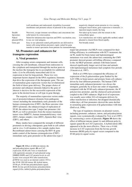
Figure 10. Effect of different introns (A)<br />
and polyadenylation signals (B) on CAT<br />
expression. ELM cells were co-transfected<br />
with equimolar amounts of each plasmid using<br />
DMRIE:DOPE and CAT protein levels in cell<br />
lysates were assayed 48 h after transfection;<br />
pCMVβ was used as an internal control. SVI is<br />
the SV40 19S/16S intron; HI, hybrid intron,<br />
SV40 pA, SV40 late polyadenylation signal;<br />
BGH, bovine growth hormone polyadenylation<br />
signal; β-Glo, rabbit β-globin polyadenylation<br />
Doll et al (1996) have compared the efficiency of<br />
expression of the β-galactosidase gene flanked by the<br />
AAV ITRs in brain tumors and primary brain cell cultures<br />
driven by four different promoters. The human CMV<br />
immediate-early enhancer/promoter was always the<br />
strongest, generally by at least one order of magnitude,<br />
compared with the SV40 early enhancer/promoter, the JC<br />
polymovirus promoter, and the chicken β-actin promoter<br />
coupled to the CMV enhancer. High level of expression<br />
was usually seen within 24 h of transgene delivery by<br />
either transfection or infection, but dropped dramatically<br />
within days; all four promoters showed the same decline<br />
in sustaining gene expression of β-galactosidase with time<br />
(Doll et al, 1996).<br />
The type of regulatory elements on plasmid vectors,<br />
including promoter, enhancer, intron, and polyadenylation<br />
signals, were systematically evaluated by Yew et al (1997)<br />
by constructing a series of plasmids. Figure 10 shows the<br />
effect of different introns (panel A) and different poly(A)<br />
signals (panel B) on CAT expression. A hybrid intron (HI)<br />
appeared to be the most effective. There was a 4-fold<br />
increase in CAT expression from the bovine growth<br />
hormone (BGH) poly(A) signal vector compared to the<br />
SV40 poly(A) signal vector.


