

01. Gene therapy Boulikas.pdf - Gene therapy & Molecular Biology
01. Gene therapy Boulikas.pdf - Gene therapy & Molecular Biology
01. Gene therapy Boulikas.pdf - Gene therapy & Molecular Biology

Create successful ePaper yourself
Turn your PDF publications into a flip-book with our unique Google optimized e-Paper software.
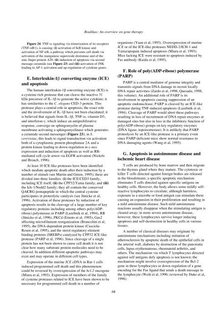
Figure 24. TNF-α signaling via trimerization of its receptors<br />
(TNF-αR1), is causing: (i) activation of IκB kinase and<br />
activation of NF-κB, a pathway which prevents cell death via<br />
activation of the manganese superoxide dismutase and of the<br />
zinc finger protein A20. (ii) induction of apoptosis via second<br />
message ceramide (see Figure 23) and (iii) activation of JNK<br />
leading to AP-1 activation and up-regulation of cytokine genes.<br />
E. Interleukin-1β converting enzyme (ICE)<br />
and apoptosis<br />
The human interleukin-1β converting enzyme (ICE) is<br />
a cysteine-rich protease that can cleave the inactive 31<br />
kDa precursor of IL-1β to generate the active cytokine; it<br />
has similarities to the C. elegans CED-3 protein. This<br />
protease plays a central role in apoptosis; the exact role<br />
and the involvement of IL-1β have not been elucidated; it<br />
is believed that signals from IL-1β, TNF-α, vitamin D3,<br />
and interferon-γ, which induce an antiproliferative<br />
response, converge on sphingomyelin of plasma<br />
membrane activating a sphingomyelinase which generates<br />
a ceramide second messenger (Figure 23); in S.<br />
cerevisiae, this leads to signal transduction via activation<br />
both of a cytoplasmic protein phosphatase 2A and a<br />
protein kinase leading to down-regulation in c-myc<br />
expression and induction of apoptosis as well as RBmediated<br />
cell cycle arrest via EGFR activation (Nickels<br />
and Broach, 1996).<br />
At least 10 ICE-like proteases have been identified<br />
which mediate apoptotic death after their induction by a<br />
number of stimuli (see Martin and Green, 1995); these are<br />
divided into three families: (i) the ICE/CED3 family,<br />
including ICE itself; (ii) the CPP32/Yama family; and (iii)<br />
the Ich-1/Nedd2 family; they all contain the conserved<br />
QACRG pentapeptide in which the central cysteine<br />
participates in proteolytic catalysis (see Jänicke et al,<br />
1996). Activation of these proteases by induction of<br />
apoptosis results in the cleavage of a large number of key<br />
regulatory proteins including among others poly(ADPribose)<br />
polymerase or PARP (Lazebnik et al, 1994), RB<br />
(Jänicke et al, 1996), PKCd (Emoto et al, 1995), Gas2<br />
affecting microfilament reorganization (Brancolini et al,<br />
1995), the DNA-dependent protein kinase (Casciola-<br />
Rosen et al, 1995), and the sterol regulatory element<br />
binding proteins (SREBPs) catalyzed by CPP32 ICE-like<br />
protease (PARP et al, 1996). Since cleavage of a single<br />
protein has not been shown to cause cell death it is not<br />
clear how many substrate protein molecules need to be<br />
cleaved. In addition different apoptotic pathways may<br />
exist and may operate in different cell types.<br />
Expression of the murine ICE cDNA in Rat-1 cells<br />
induced programmed cell death and this phenomenon<br />
could be reversed by overexpression of the bcl-2 oncogene<br />
(Miura et al, 1993). Expression of members of the family<br />
of cysteine proteases related to ICE have been shown to be<br />
necessary for programmed cell death in a number of<br />
<strong>Boulikas</strong>: An overview on gene <strong>therapy</strong><br />
68<br />
organisms (Yuan et al, 1993). Overexpression of murine<br />
ICE or of the ICE-like proteases NEDD-2/ICH-1 and<br />
Yama/apopain induced apoptosis (Miura et al, 1993).<br />
Mice lacking ICE were resistant to apoptosis induced by<br />
Fas antibody (Kuida et al, 1995).<br />
F. Role of poly(ADP-ribose) polymerase<br />
(PARP)<br />
PARP is a central mediator of genome integrity and<br />
transmits signals from DNA damage to recruit locally<br />
DNA repair activities (Zardo et al, 1998; Quesada, 1998,<br />
this volume). An additional role of PARP is its<br />
involvement in apoptosis causing suppression of an<br />
apoptotic endonuclease; PARP is cleaved by an ICE-like<br />
protease during TNF-induced apoptosis (Lazebnik et al,<br />
1994). Cleavage of PARP would abort these pathways<br />
resulting in loss of recruitment of DNA repair enzymes at<br />
damaged sites but also in loss in the inhibitory function of<br />
poly(ADP-ribose) groups on key regulatory enzymes<br />
(DNA ligase, topoisomerase). It is unlikely that PARP<br />
proteolysis by an ICE-like protease is a primary event<br />
since PARP-deficient mice show normal resistance to<br />
DNA damaging agents (Wang et al, 1995).<br />
G. Apoptosis in autoimmune disease and<br />
ischemic heart disease<br />
T cells are produced by bone marrow and then migrate<br />
to the thymus gland where they mature. The cytotoxic or<br />
killer T cells directed against foreign bodies are released<br />
in the bloodstream; a specific apoptotic mechanism<br />
eliminates T cells directed against specific antigens on<br />
healthy cells. However, the body allows some mildly selfreactive<br />
lymphocytes to circulate; although harmless,<br />
exposure to a microbe or food antigen can stimulate them<br />
causing an expansion in their proliferation and resulting in<br />
a mild autoimmune disease. Such mild autoimmune<br />
reactions usually disappear when the stimulating antigen is<br />
cleared away; in more severe autoimmune disease,<br />
however, these lymphocytes survive longer inducing<br />
apoptosis and self-destruction in healthy cells in various<br />
tissues.<br />
A number of classical diseases may originate by<br />
autoimmune mechanisms including initiation of<br />
atherosclerosis by apoptotic death of the epithelial cells in<br />
the arterial wall, diabetes by destruction of the pancreatic<br />
cells, lupus erythematosus, rheumatoid arthritis, and<br />
others. The mechanism via which T lymphocytes directed<br />
against self antigens defy apoptosis is not known; the<br />
mechanism might involve overexpression of the Bcl-2<br />
gene in these lymphocytes or down-regulation of a gene<br />
encoding for the Fas ligand that sends a death message to<br />
the lymphocyte (Weih et al, 1996; reviewed by Duke et al,<br />
1996).


