

01. Gene therapy Boulikas.pdf - Gene therapy & Molecular Biology
01. Gene therapy Boulikas.pdf - Gene therapy & Molecular Biology
01. Gene therapy Boulikas.pdf - Gene therapy & Molecular Biology

You also want an ePaper? Increase the reach of your titles
YUMPU automatically turns print PDFs into web optimized ePapers that Google loves.
<strong>Gene</strong> Therapy and <strong>Molecular</strong> <strong>Biology</strong> Vol 1, page 55<br />
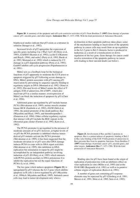
Figure 20. A summary of the apoptotic and cell cycle restrictive activities of p53. From <strong>Boulikas</strong> T (1997) <strong>Gene</strong> <strong>therapy</strong> of prostate<br />
cancer: p53, suicidal genes, and other targets. Anticancer Res 17, 1471-1506. With the kind permission of Anticancer Research.<br />
biophysical studies indicate that p53 exists as a tetramer in<br />
solution (Stenger et al., 1992).<br />
Increased levels of p53 upregulate the expression of<br />
specific genes including Cip-1/Waf-1/p21 (El-Deiry et al,<br />
1993), GADD45 (Kastan et al, 1992), cyclin G (Okamoto<br />
and Beach, 1994), and mdm2 (Perry et al, 1993; Barak et<br />
al, 1993; Momand et al, 1992) which is induced by UV<br />
damage in a p53-dependent pathway (Perry et al, 1993).<br />
Gadd45 inhibits cell cycle progression (Papathanasiou et<br />
al, 1991).<br />
Mdm2 acts as a feedback loop for the biological<br />
functions of p53 apparently to moderate the G1/S arrest or<br />
apoptosis triggered by p53 following severe damage to<br />
DNA. Mdm2 protein associates with p53 causing p53<br />
inactivation by preventing its sequence-specific binding to<br />
regulatory targets in DNA (Momand et al, 1992; Oliner et<br />
al, 1992). Elevated levels of Mdm2 mimic the effect of T<br />
antigen, E1B of adenovirus, E6 of HPV, which also<br />
inactivate p53 in a similar manner; overexpression of<br />
Mdm2 can block the induction of apoptosis by p53 (Chen<br />
et al, 1994).<br />
Additional genes up-regulated by p53 include human<br />
PCNA (Shivakumar et al, 1995), mouse muscle creatine<br />
kinase MCK (Zambetti et al, 1992), EGFR (Deb et al,<br />
1994), the potent promoter of the death pathway Bax<br />
(Miyashita and Reed, 1995), and thrombospondin-1<br />
(Dameron et al, 1994). Other cellular regulatory regions<br />
that interact with p53 include the RGC repeats in the<br />
ribosomal gene cluster (Farmer et al, 1992; Kern et al,<br />
1992).<br />
The PCNA promoter is up-regulated in the presence of<br />
moderate amounts of wt p53; however, at higher levels of<br />
wt p53 the PCNA promoter is inhibited whereas tumorderived<br />
p53 mutants activate the PCNA promoter<br />
(Shivakumar et al, 1995); it has been suggested that the<br />
moderate elevation in wt p53 seen after DNA damage<br />
induces PCNA to cope with its DNA repair activities<br />
(Shivakumar et al, 1995); this inhibition in DNA<br />
replication but stimulation in repair by p53 might be<br />
accomplished by an independent pathway involving<br />
induction of p21 (El-Deiry et al, 1993) which interacts<br />
with PCNA protein auxiliary to DNA polymerase δ to<br />
inhibit the replication but not the repair functions of<br />
PCNA (Li et al, 1994).<br />
The bax gene which induces apoptosis (Figure 21) is<br />
upregulated by p53 whereas the bcl-2 gene which inhibits<br />
apoptosis in B cells is down-regulated by p53 (Miyashita<br />
et al, 1994a,b; Miyashita and Reed, 1995). Initiated cancer<br />
cells may lead to tumor development only when a<br />
55<br />
dysfunction in their apoptotic pathway takes place; some<br />
of the mechanisms leading to inactivation of the apoptotic<br />
pathway in cancer cells may result from an up-regulation<br />
in the bcl-2 gene (a Bcl-2 chimeric factor is produced in<br />
leukemias as a result of a translocation) or downregulation<br />
of the bax gene. <strong>Gene</strong> <strong>therapy</strong> for cancer could<br />
involve restoration of the apoptotic pathway in cancer<br />
cells leading to their suicidal death (see below).<br />
Figure 21. Involvement of Bax and Bcl-2 proteins in<br />
apoptosis. Bax is a potent inducer of apoptosis; binding of Bcl-2<br />
to Bax (also binding of the E1B 19 kDa protein of adenovirus to<br />
Bax) prevents Bax from its apoptotic functions. From <strong>Boulikas</strong> T<br />
(1997) <strong>Gene</strong> <strong>therapy</strong> of prostate cancer: p53, suicidal genes, and<br />
other targets. Anticancer Res 17, 1471-1506. With the kind<br />
permission of Anticancer Research.<br />
Binding sites for p53 have been found at the origin of<br />
replication of polyomavirus with an inhibitory effect on<br />
virus replication in vitro (Miller et al, 1995) and at the<br />
SV40 ORI (Bargonetti et al, 1991) as well as in putative<br />
cellular origins of replication (Kern et al, 1991).<br />
A number of genes not containing p53 response<br />
elements may be repressed by p53 (Ginsberg et al, 1991;<br />
Mercer et al, 1991; Shiio et al, 1992; Seto et al, 1992).


