

01. Gene therapy Boulikas.pdf - Gene therapy & Molecular Biology
01. Gene therapy Boulikas.pdf - Gene therapy & Molecular Biology
01. Gene therapy Boulikas.pdf - Gene therapy & Molecular Biology

Create successful ePaper yourself
Turn your PDF publications into a flip-book with our unique Google optimized e-Paper software.
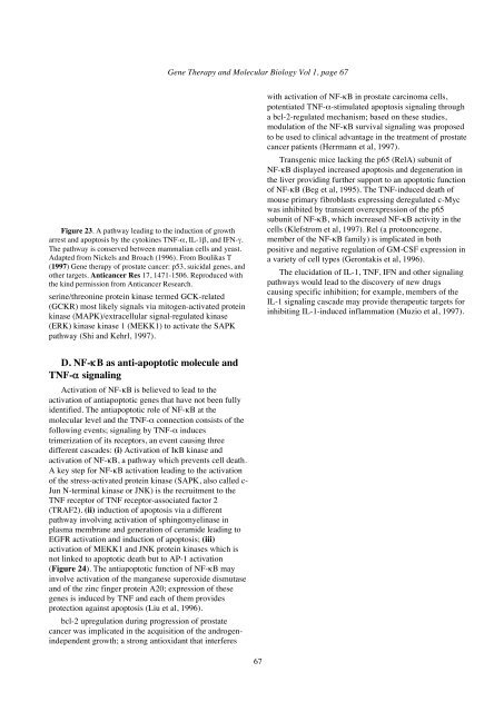
Figure 23. A pathway leading to the induction of growth<br />
arrest and apoptosis by the cytokines TNF-α, IL-1β, and IFN-γ.<br />
The pathway is conserved between mammalian cells and yeast.<br />
Adapted from Nickels and Broach (1996). From <strong>Boulikas</strong> T<br />
(1997) <strong>Gene</strong> <strong>therapy</strong> of prostate cancer: p53, suicidal genes, and<br />
other targets. Anticancer Res 17, 1471-1506. Reproduced with<br />
the kind permission from Anticancer Research.<br />
serine/threonine protein kinase termed GCK-related<br />
(GCKR) most likely signals via mitogen-activated protein<br />
kinase (MAPK)/extracellular signal-regulated kinase<br />
(ERK) kinase kinase 1 (MEKK1) to activate the SAPK<br />
pathway (Shi and Kehrl, 1997).<br />
D. NF-κB as anti-apoptotic molecule and<br />
TNF-α signaling<br />
Activation of NF-κB is believed to lead to the<br />
activation of antiapoptotic genes that have not been fully<br />
identified. The antiapoptotic role of NF-κB at the<br />
molecular level and the TNF-α connection consists of the<br />
following events; signaling by TNF-α induces<br />
trimerization of its receptors, an event causing three<br />
different cascades: (i) Activation of IκB kinase and<br />
activation of NF-κB, a pathway which prevents cell death.<br />
A key step for NF-κB activation leading to the activation<br />
of the stress-activated protein kinase (SAPK, also called c-<br />
Jun N-terminal kinase or JNK) is the recruitment to the<br />
TNF receptor of TNF receptor-associated factor 2<br />
(TRAF2). (ii) induction of apoptosis via a different<br />
pathway involving activation of sphingomyelinase in<br />
plasma membrane and generation of ceramide leading to<br />
EGFR activation and induction of apoptosis; (iii)<br />
activation of MEKK1 and JNK protein kinases which is<br />
not linked to apoptotic death but to AP-1 activation<br />
(Figure 24). The antiapoptotic function of NF-κB may<br />
involve activation of the manganese superoxide dismutase<br />
and of the zinc finger protein A20; expression of these<br />
genes is induced by TNF and each of them provides<br />
protection against apoptosis (Liu et al, 1996).<br />
bcl-2 upregulation during progression of prostate<br />
cancer was implicated in the acquisition of the androgenindependent<br />
growth; a strong antioxidant that interferes<br />
<strong>Gene</strong> Therapy and <strong>Molecular</strong> <strong>Biology</strong> Vol 1, page 67<br />
67<br />
with activation of NF-κB in prostate carcinoma cells,<br />
potentiated TNF-α-stimulated apoptosis signaling through<br />
a bcl-2-regulated mechanism; based on these studies,<br />
modulation of the NF-κB survival signaling was proposed<br />
to be used to clinical advantage in the treatment of prostate<br />
cancer patients (Herrmann et al, 1997).<br />
Transgenic mice lacking the p65 (RelA) subunit of<br />
NF-κB displayed increased apoptosis and degeneration in<br />
the liver providing further support to an apoptotic function<br />
of NF-κB (Beg et al, 1995). The TNF-induced death of<br />
mouse primary fibroblasts expressing deregulated c-Myc<br />
was inhibited by transient overexpression of the p65<br />
subunit of NF-κB, which increased NF-κB activity in the<br />
cells (Klefstrom et al, 1997). Rel (a protooncogene,<br />
member of the NF-κB family) is implicated in both<br />
positive and negative regulation of GM-CSF expression in<br />
a variety of cell types (Gerontakis et al, 1996).<br />
The elucidation of IL-1, TNF, IFN and other signaling<br />
pathways would lead to the discovery of new drugs<br />
causing specific inhibition; for example, members of the<br />
IL-1 signaling cascade may provide therapeutic targets for<br />
inhibiting IL-1-induced inflammation (Muzio et al, 1997).


