01. Gene therapy Boulikas.pdf - Gene therapy & Molecular Biology
01. Gene therapy Boulikas.pdf - Gene therapy & Molecular Biology
01. Gene therapy Boulikas.pdf - Gene therapy & Molecular Biology
You also want an ePaper? Increase the reach of your titles
YUMPU automatically turns print PDFs into web optimized ePapers that Google loves.
<strong>Boulikas</strong>: An overview on gene <strong>therapy</strong><br />
78<br />
Li et al, 1994; Zhao et al, 1997<br />
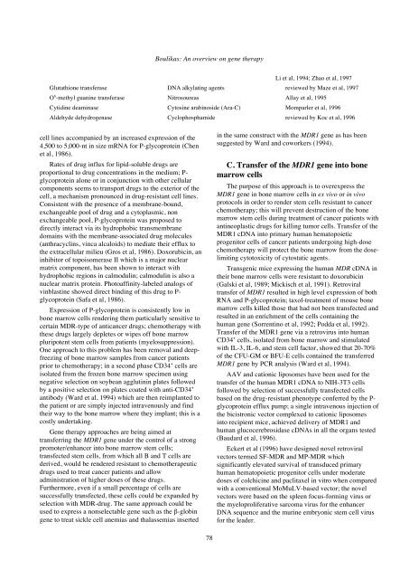
Glutathione transferase DNA alkylating agents reviewed by Maze et al, 1997<br />
O 6 -methyl guanine transferase Nitrosoureas Allay et al, 1995<br />
Cytidine deaminase Cytosine arabinoside (Ara-C) Momparler et al, 1996<br />
Aldehyde dehydrogenase Cyclophosphamide reviewed by Koc et al, 1996<br />
cell lines accompanied by an increased expression of the<br />
4,500 to 5,000-nt in size mRNA for P-glycoprotein (Chen<br />
et al, 1986).<br />
Rates of drug influx for lipid-soluble drugs are<br />
proportional to drug concentrations in the medium; Pglycoprotein<br />
alone or in conjunction with other cellular<br />
components seems to transport drugs to the exterior of the<br />
cell, a mechanism pronounced in drug-resistant cell lines.<br />
Consistent with the presence of a membrane-bound,<br />
exchangeable pool of drug and a cytoplasmic, non<br />
exchangeable pool, P-glycoprotein was proposed to<br />
directly interact via its hydrophobic transmembrane<br />
domains with the membrane-associated drug molecules<br />
(anthracyclins, vinca alcaloids) to mediate their efflux to<br />
the extracellular milieu (Gros et al, 1986). Doxorubicin, an<br />
inhibitor of topoisomerase II which is a major nuclear<br />
matrix component, has been shown to interact with<br />
hydrophobic regions in calmodulin; calmodulin is also a<br />
nuclear matrix protein. Photoaffinity-labeled analogs of<br />
vinblastine showed direct binding of this drug to Pglycoprotein<br />
(Safa et al, 1986).<br />
Expression of P-glycoprotein is consistently low in<br />
bone marrow cells rendering them particularly sensitive to<br />
certain MDR-type of anticancer drugs; chemo<strong>therapy</strong> with<br />
these drugs largely depletes or wipes off bone marrow<br />
pluripotent stem cells from patients (myelosuppression).<br />
One approach to this problem has been removal and deepfreezing<br />
of bone marrow samples from cancer patients<br />
prior to chemo<strong>therapy</strong>; in a second phase CD34 + cells are<br />
isolated from the frozen bone marrow specimen using<br />
negative selection on soybean agglutinin plates followed<br />
by a positive selection on plates coated with anti-CD34 +<br />
antibody (Ward et al, 1994) which are then reimplanted to<br />
the patient or are simply injected intravenously and find<br />
their way to the bone marrow where they implant; this is a<br />
costly undertaking.<br />
<strong>Gene</strong> <strong>therapy</strong> approaches are being aimed at<br />
transferring the MDR1 gene under the control of a strong<br />
promoter/enhancer into bone marrow stem cells;<br />
transfected stem cells, from which all B and T cells are<br />
derived, would be rendered resistant to chemotherapeutic<br />
drugs used to treat cancer patients and allow<br />
administration of higher doses of these drugs.<br />
Furthermore, even if a small percentage of cells are<br />
successfully transfected, these cells could be expanded by<br />
selection with MDR-drug. The same approach could be<br />
used to express a nonselectable gene such as the β-globin<br />
gene to treat sickle cell anemias and thalassemias inserted<br />
in the same construct with the MDR1 gene as has been<br />
suggested by Ward and coworkers (1994).<br />
C. Transfer of the MDR1 gene into bone<br />
marrow cells<br />
The purpose of this approach is to overexpress the<br />
MDR1 gene in bone marrow cells in ex vivo or in vivo<br />
protocols in order to render stem cells resistant to cancer<br />
chemo<strong>therapy</strong>; this will prevent destruction of the bone<br />
marrow stem cells during treatment of cancer patients with<br />
antineoplastic drugs for killing tumor cells. Transfer of the<br />
MDR1 cDNA into primary human hematopoietic<br />
progenitor cells of cancer patients undergoing high-dose<br />
chemo<strong>therapy</strong> will protect the bone marrow from the doselimiting<br />
cytotoxicity of cytostatic agents.<br />
Transgenic mice expressing the human MDR cDNA in<br />
their bone marrow cells were resistant to doxorubicin<br />
(Galski et al, 1989; Mickisch et al, 1991). Retroviral<br />
transfer of MDR1 resulted in high level expression of both<br />
RNA and P-glycoprotein; taxol-treatment of mouse bone<br />
marrow cells killed those that had not been transfected and<br />
resulted in an enrichment of the cells containing the<br />
human gene (Sorrentino et al, 1992; Podda et al, 1992).<br />
Transfer of the MDR1 gene via a retrovirus into human<br />
CD34 + cells, isolated from bone marrow and stimulated<br />
with IL-3, IL-6, and stem cell factor, showed that 20-70%<br />
of the CFU-GM or BFU-E cells contained the transferred<br />
MDR1 gene by PCR analysis (Ward et al, 1994).<br />
AAV and cationic liposomes have been used for the<br />
transfer of the human MDR1 cDNA to NIH-3T3 cells<br />
followed by selection of successfully transfected cells<br />
based on the drug-resistant phenotype conferred by the Pglycoprotein<br />
efflux pump; a single intravenous injection of<br />
the bicistronic vector complexed to cationic liposomes<br />
into recipient mice, achieved delivery of MDR1 and<br />
human glucocerebrosidase cDNAs in all the organs tested<br />
(Baudard et al, 1996).<br />
Eckert et al (1996) have designed novel retroviral<br />
vectors termed SF-MDR and MP-MDR which<br />
significantly elevated survival of transduced primary<br />
human hematopoietic progenitor cells under moderate<br />
doses of colchicine and paclitaxel in vitro when compared<br />
with a conventional MoMuLV-based vector; the novel<br />
vectors were based on the spleen focus-forming virus or<br />
the myeloproliferative sarcoma virus for the enhancer<br />
DNA sequence and the murine embryonic stem cell virus<br />
for the leader.


