

01. Gene therapy Boulikas.pdf - Gene therapy & Molecular Biology
01. Gene therapy Boulikas.pdf - Gene therapy & Molecular Biology
01. Gene therapy Boulikas.pdf - Gene therapy & Molecular Biology

You also want an ePaper? Increase the reach of your titles
YUMPU automatically turns print PDFs into web optimized ePapers that Google loves.
<strong>Gene</strong> Therapy and <strong>Molecular</strong> <strong>Biology</strong> Vol 1, page 77<br />
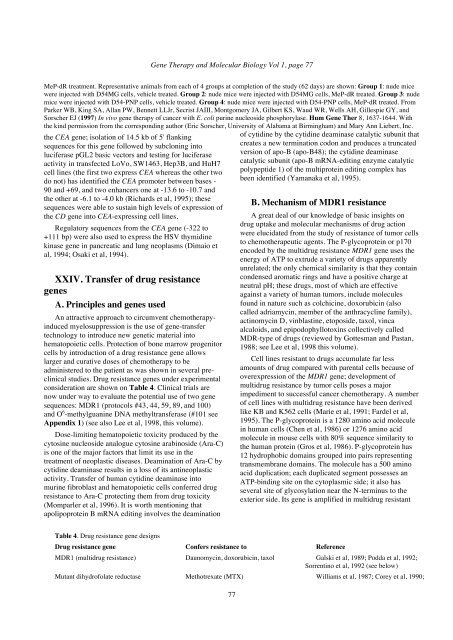
MeP-dR treatment. Representative animals from each of 4 groups at completion of the study (62 days) are shown: Group 1: nude mice<br />
were injected with D54MG cells, vehicle treated. Group 2: nude mice were injected with D54MG cells, MeP-dR treated. Group 3: nude<br />
mice were injected with D54-PNP cells, vehicle treated. Group 4: nude mice were injected with D54-PNP cells, MeP-dR treated. From<br />
Parker WB, King SA, Allan PW, Bennett LLJr, Secrist JAIII, Montgomery JA, Gilbert KS, Waud WR, Wells AH, Gillespie GY, and<br />
Sorscher EJ (1997) In vivo gene <strong>therapy</strong> of cancer with E. coli purine nucleoside phosphorylase. Hum <strong>Gene</strong> Ther 8, 1637-1644. With<br />
the kind permission from the corresponding author (Eric Sorscher, University of Alabama at Birmingham) and Mary Ann Liebert, Inc.<br />
the CEA gene; isolation of 14.5 kb of 5' flanking<br />
sequences for this gene followed by subcloning into<br />
luciferase pGL2 basic vectors and testing for luciferase<br />
activity in transfected LoVo, SW1463, Hep3B, and HuH7<br />
cell lines (the first two express CEA whereas the other two<br />
do not) has identified the CEA promoter between bases -<br />
90 and +69, and two enhancers one at -13.6 to -10.7 and<br />
the other at -6.1 to -4.0 kb (Richards et al, 1995); these<br />
sequences were able to sustain high levels of expression of<br />
the CD gene into CEA-expressing cell lines.<br />
Regulatory sequences from the CEA gene (-322 to<br />
+111 bp) were also used to express the HSV thymidine<br />
kinase gene in pancreatic and lung neoplasms (Dimaio et<br />
al, 1994; Osaki et al, 1994).<br />
XXIV. Transfer of drug resistance<br />
genes<br />
A. Principles and genes used<br />
An attractive approach to circumvent chemo<strong>therapy</strong>induced<br />
myelosuppression is the use of gene-transfer<br />
technology to introduce new genetic material into<br />
hematopoietic cells. Protection of bone marrow progenitor<br />
cells by introduction of a drug resistance gene allows<br />
larger and curative doses of chemo<strong>therapy</strong> to be<br />
administered to the patient as was shown in several preclinical<br />
studies. Drug resistance genes under experimental<br />
consideration are shown on Table 4. Clinical trials are<br />
now under way to evaluate the potential use of two gene<br />
sequences: MDR1 (protocols #43, 44, 59, 89, and 100)<br />
and O 6 -methylguanine DNA methyltransferase (#101 see<br />
Appendix 1) (see also Lee et al, 1998, this volume).<br />
Dose-limiting hematopoietic toxicity produced by the<br />
cytosine nucleoside analogue cytosine arabinoside (Ara-C)<br />
is one of the major factors that limit its use in the<br />
treatment of neoplastic diseases. Deamination of Ara-C by<br />
cytidine deaminase results in a loss of its antineoplastic<br />
activity. Transfer of human cytidine deaminase into<br />
murine fibroblast and hematopoietic cells conferred drug<br />
resistance to Ara-C protecting them from drug toxicity<br />
(Momparler et al, 1996). It is worth mentioning that<br />
apolipoprotein B mRNA editing involves the deamination<br />
Table 4. Drug resistance gene designs<br />
77<br />
of cytidine by the cytidine deaminase catalytic subunit that<br />
creates a new termination codon and produces a truncated<br />
version of apo-B (apo-B48); the cytidine deaminase<br />
catalytic subunit (apo-B mRNA-editing enzyme catalytic<br />
polypeptide 1) of the multiprotein editing complex has<br />
been identified (Yamanaka et al, 1995).<br />
B. Mechanism of MDR1 resistance<br />
A great deal of our knowledge of basic insights on<br />
drug uptake and molecular mechanisms of drug action<br />
were elucidated from the study of resistance of tumor cells<br />
to chemotherapeutic agents. The P-glycoprotein or p170<br />
encoded by the multidrug resistance MDR1 gene uses the<br />
energy of ATP to extrude a variety of drugs apparently<br />
unrelated; the only chemical similarity is that they contain<br />
condensed aromatic rings and have a positive charge at<br />
neutral pH; these drugs, most of which are effective<br />
against a variety of human tumors, include molecules<br />
found in nature such as colchicine, doxorubicin (also<br />
called adriamycin, member of the anthracycline family),<br />
actinomycin D, vinblastine, etoposide, taxol, vinca<br />
alcaloids, and epipodophyllotoxins collectively called<br />
MDR-type of drugs (reviewed by Gottesman and Pastan,<br />
1988; see Lee et al, 1998 this volume).<br />
Cell lines resistant to drugs accumulate far less<br />
amounts of drug compared with parental cells because of<br />
overexpression of the MDR1 gene; development of<br />
multidrug resistance by tumor cells poses a major<br />
impediment to successful cancer chemo<strong>therapy</strong>. A number<br />
of cell lines with multidrug resistance have been derived<br />
like KB and K562 cells (Marie et al, 1991; Fardel et al,<br />
1995). The P-glycoprotein is a 1280 amino acid molecule<br />
in human cells (Chen et al, 1986) or 1276 amino acid<br />
molecule in mouse cells with 80% sequence similarity to<br />
the human protein (Gros et al, 1986). P-glycoprotein has<br />
12 hydrophobic domains grouped into pairs representing<br />
transmembrane domains. The molecule has a 500 amino<br />
acid duplication; each duplicated segment possesses an<br />
ATP-binding site on the cytoplasmic side; it also has<br />
several site of glycosylation near the N-terminus to the<br />
exterior side. Its gene is amplified in multidrug resistant<br />
Drug resistance gene Confers resistance to Reference<br />
MDR1 (multidrug resistance) Daunomycin, doxorubicin, taxol Galski et al, 1989; Podda et al, 1992;<br />
Sorrentino et al, 1992 (see below)<br />
Mutant dihydrofolate reductase Methotrexate (MTX) Williams et al, 1987; Corey et al, 1990;


