DRAFT Recommended Practice for Measurements and ...
DRAFT Recommended Practice for Measurements and ...
DRAFT Recommended Practice for Measurements and ...

You also want an ePaper? Increase the reach of your titles
YUMPU automatically turns print PDFs into web optimized ePapers that Google loves.
1/29/98 121 C95.3-1991 Revision — 2 nd Draft<br />
10/98 Draft<br />
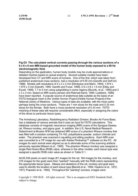
Fig D3 The calculated vertical currents passing through the various sections of a<br />
6 x 6 x 6 mm MRI-based grounded model of the human body exposed to a 60 Hz<br />
electromagnetic field.<br />
Depending on the application, human body models may be crude approximations or<br />
detailed meshes based on actual anatomy. Several suitable models have been<br />
developed from CT <strong>and</strong> MRI scans of humans. One of the first, which was taken from<br />
published anatomical cross sections, had a resolution of 6.55 mm [G<strong>and</strong>hi <strong>and</strong> DeFord,<br />
1988]. Models with resolutions of 2 x 2 x 2 mm [Dimbylow <strong>and</strong> Mann, 1994], 1.974 x<br />
1.974 x 3 mm [G<strong>and</strong>hi, 1995; G<strong>and</strong>hi <strong>and</strong> Furse, 1995], 0.9 x 0.9 x 1.5 mm [Olley <strong>and</strong><br />
Excell, 1995], 1.7 to 5 mm using subgridding in some regions [Stuchly, et al., 1995] <strong>and</strong> 3<br />
x 3 x 3 mm, based on MRI scans [Jensen <strong>and</strong> Rahmat-Samii, 1995; Luebbers, 1996]<br />
have been reported. A popular source of anatomical data suitable as the basis of an<br />
FDTD biological mesh is the Visible Human Project [Visible Human Project] of the<br />
National Library of Medicine. Various types of data are available, with the most useful<br />
perhaps being the cross sections. These are 1 mm slices <strong>for</strong> the male <strong>and</strong> 0.33 mm<br />
slices <strong>for</strong> the female. Both have a cross-sectional resolution of 0.33 mm. FDTD<br />
meshing of these data still requires considerable ef<strong>for</strong>t, especially in assigning the colors<br />
of the slices to particular tissue types.<br />
The Armstrong Laboratory, Radiofrequency Radiation Division, Brooks Air Force Base,<br />
has a database of various animals that it uses as input <strong>for</strong> FDTD calculations. This<br />
database consists of magnetic resonance imaging (MRI) scans of the Sprague-Dawley<br />
rat, Rhesus monkey, <strong>and</strong> pigmy goat. In addition, the U.S. Naval Medical Research<br />
Detachment at Brooks AFB has obtained MRI scans of a phantom Rhesus monkey that<br />
was filled with a solution containing TX-150, polyethylene powder, sodium chloride <strong>and</strong><br />
water. The phantom was scanned in squatting <strong>and</strong> sitting positions. Each MRI scan<br />
was converted to a TIF image <strong>for</strong> use with Adobe Photoshop software on the PC. All<br />
images <strong>for</strong> each animal were aligned so as to eliminate some of the scanning artifacts<br />
previously reported [Mason et al., 1995]. The phantom Rhesus monkey was assigned a<br />
single Red-Green-Blue (RGB) value, whereas in the other models, each tissue type was<br />
assigned a specific RGB color value as shown in Table D 1.<br />
All 65,536 pixels on each image (81 images <strong>for</strong> the rat, 184 images <strong>for</strong> the monkey, <strong>and</strong><br />
276 images <strong>for</strong> the goat) were then "painted" manually with the RGB colors representing<br />
the appropriate tissue types. Atlases <strong>and</strong> skeletons of the rat, monkey <strong>and</strong> goat were<br />
used to identify the location of each tissue type [Barrett et al., 1994; Hopkins et al., 1972,<br />
1973; Popesko et al., 1992]. Throughout the "painting" process, images were<br />
Copyright © 1998 IEEE. All rights reserved. This is an unapproved IEEE St<strong>and</strong>ards Draft,<br />
subject to change.