DRAFT Recommended Practice for Measurements and ...
DRAFT Recommended Practice for Measurements and ...
DRAFT Recommended Practice for Measurements and ...

Create successful ePaper yourself
Turn your PDF publications into a flip-book with our unique Google optimized e-Paper software.
1/29/98 66 C95.3-1991 Revision — 2 nd Draft<br />
10/98 Draft<br />
spheres [B15, B116] <strong>and</strong> in waveguides filled with lossy dielectric liquids, e.g. salt water,<br />
[B59]. These lossy media are exposed to a known external E-field <strong>and</strong> then placed in the<br />
dielectric object in which the internal E-field distribution has been calculated using<br />
electromagnetic field theory. The internal E-field calibration factor <strong>for</strong> the probe is<br />
determined from its response compared with the calculated internal field. If this is done<br />
at several frequencies <strong>for</strong> a specific probe design, the calibration uncertainty will be<br />
typically 1 to 2 dB when implanted in any lossy, high dielectric-constant object, such as<br />
biological tissue that contains a large percentage of water (muscle, brain, <strong>and</strong> internal<br />
organs, but not bone or fat). (See also 5.5.2.)<br />
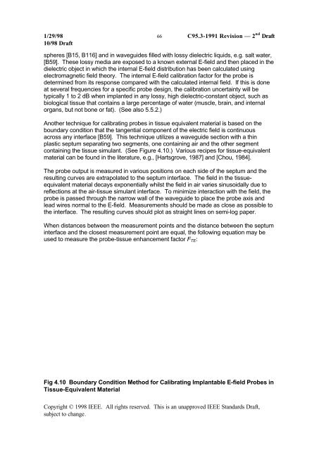
Another technique <strong>for</strong> calibrating probes in tissue equivalent material is based on the<br />
boundary condition that the tangential component of the electric field is continuous<br />
across any interface [B59]. This technique utilizes a waveguide section with a thin<br />
plastic septum separating two segments, one containing air <strong>and</strong> the other segment<br />
containing the tissue simulant. (See Figure 4.10.) Various recipes <strong>for</strong> tissue-equivalent<br />
material can be found in the literature, e.g., [Hartsgrove, 1987] <strong>and</strong> [Chou, 1984].<br />
The probe output is measured in various positions on each side of the septum <strong>and</strong> the<br />
resulting curves are extrapolated to the septum interface. The field in the tissueequivalent<br />
material decays exponentially whilst the field in air varies sinusoidally due to<br />
reflections at the air-tissue simulant interface. To minimize interaction with the field, the<br />
probe is passed through the narrow wall of the waveguide to place the probe axis <strong>and</strong><br />
lead wires normal to the E-field. <strong>Measurements</strong> should be made as close as possible to<br />
the interface. The resulting curves should plot as straight lines on semi-log paper.<br />
When distances between the measurement points <strong>and</strong> the distance between the septum<br />
interface <strong>and</strong> the closest measurement point are equal, the following equation may be<br />
used to measure the probe-tissue enhancement factor F TE :<br />
Fig 4.10 Boundary Condition Method <strong>for</strong> Calibrating Implantable E-field Probes in<br />
Tissue-Equivalent Material<br />
Copyright © 1998 IEEE. All rights reserved. This is an unapproved IEEE St<strong>and</strong>ards Draft,<br />
subject to change.