- Page 2 and 3:
SEER Program Self InstructionalManu
- Page 4 and 5:
TABLE OF CONTENTS BOOK 5: ABSTRACTI
- Page 6:
SECTION A OBJECTIVES AND CONTENT OF
- Page 10:
SECTION B THE COMPOSITION AND ORGAN
- Page 13 and 14:
Physical Examination General (gener
- Page 15 and 16:
FORMS USED TO RECORD INFORMATION IN
- Page 18:
SECTION C ABSTRACTING A MEDICAL REC
- Page 21 and 22:
WHEN TO ABSTRACT CASES While it may
- Page 23 and 24:
Local Registry Number A patient's r
- Page 25 and 26:
Marital Status Select appropriate a
- Page 27 and 28:
Abstracting Diagnostic Procedures S
- Page 30 and 31:
SECTION D DIAGNOSTIC PROCEDURES: GE
- Page 32:
SECTION E CLINICAL EXAMINATIONS: PH
- Page 36:
EXAMPLE E1 Name: Sarah O. Luckless
- Page 40:
EXAMPLE E2 Name: Hector Breathless
- Page 44 and 45:
BODY SECTION RADIOGRAPHY. Body sect
- Page 46:
E. Lower GI Series or Barium Enema:
- Page 50:
Example E3 can be abstracted as fol
- Page 54:
Example E4 can be abstracted as fol
- Page 58:
Example E5 can be abstracted as fol
- Page 62:
Example E6 might be abstracted as f
- Page 66:
Example E7 can be abstracted as fol
- Page 70:
EXAMPLE E9 Name: Hardy Smooker Hosp
- Page 74:
Examples E8-E10 can be abstracted a
- Page 78:
EXAMPLE Ell Name: Emma Bronchilli H
- Page 81 and 82:
DIAGNOSTIC NUCLEAR MEDICINE EXAMINA
- Page 84:
EXAMPLE El2 THE DIVISION OF NUCLEAR
- Page 88:
EXAMPLE E14 Record No.: 000014 Name
- Page 92:
MAGNETIC RESONANCE IMAGING: Magneti
- Page 96:
EXAMPLEE16 MRI CONSULTATIONREPORT N
- Page 100 and 101:
HEMATOLOGIC EXAMINATION A hematolog
- Page 102 and 103:
Granulocytes or granular leukocytes
- Page 104 and 105:
Certain types of disease associated
- Page 106 and 107:
EXAMPLE E17 HEMATOLOGY LABORATORY N
- Page 108 and 109:
EXAMPLEE18 HEMATOLOGY LABORATORY Na
- Page 110 and 111:
EXAMPLE E19 HEMATOLOGY LABORATORY R
- Page 112 and 113:
PRACTICAL EXERCISE ANSWERS Example
- Page 114 and 115:
Bone Marrow Studies. Examination of
- Page 116 and 117:
EXAMPLE E20 Name: Bo Comeaway Hospi
- Page 118 and 119:
Example E20 can be abstracted as fo
- Page 120 and 121:
Blood Serum Studies Automation and
- Page 122 and 123:
Other Laboratory Studies Marrow Aci
- Page 124 and 125:
Totalprotein. This test measures th
- Page 126 and 127:
SECTION F MANIPULATIVE AND OPERATIV
- Page 128 and 129:
SECTION F MANIPULATIVE AND OPERATIV
- Page 130 and 131:
EXAMPLE FI Rec. No. 000019 Name Iv_
- Page 132 and 133:
The manner in which the findings in
- Page 134 and 135:
EXAMPLE F2 LARYNGOSCOPY REPORT Hosp
- Page 136 and 137:
The illustrated report can be abstr
- Page 138 and 139:
I EXAMPLE F3 REPORT OF CYSTOSCOPY N
- Page 140 and 141:
Example F3 is abstracted as follows
- Page 142 and 143:
EXAMPLE F4 GASTROINTESTINAL DIAGNOS
- Page 144 and 145:
The information described in Exampl
- Page 146 and 147:
EXAMPLE F5 SIGMOIDOSCOPY REPORT Rec
- Page 148 and 149:
EXAMPLE F6 PROCTOSCOPY REPORT Rec.
- Page 150 and 151:
EXAMPLE F7 GASTROSCOPIC REPORT Hosp
- Page 152 and 153:
EXAMPLE F8 GASTROSCOPIC REPORT Hosp
- Page 154 and 155:
EXAMPLE F9 BRONCHOSCOPY REPORT Hosp
- Page 156 and 157:
EXAMPLE FIO BRONCHOSCOPY REPORT Hos
- Page 158 and 159:
EXAMPLE Fll BRONCHOSCOPY REPORT Hos
- Page 160 and 161: EXAMPLE F12 BRONCHOSCOPY REPORT Hos
- Page 162 and 163: PRACTICAL EXERCISE ANSWERS These en
- Page 164 and 165: Example F10: 5/5/91. Bronchoscopy:
- Page 166 and 167: In all of the "oscopies" described
- Page 168 and 169: EXAMPLE F13 OPERATIVE REPORT Name:
- Page 170 and 171: This operative report may be abstra
- Page 172 and 173: OPERATIVE PROCEDURES Exploratory_Su
- Page 174 and 175: EXAMPLE F14 OPERATIVE REPORT Servic
- Page 176 and 177: The operative record used as an ill
- Page 178 and 179: EXAMPLE F15 OPERATIVE REPORT Servic
- Page 180 and 181: Example F15: 3/11/91. Expl. Lap.: L
- Page 182 and 183: PRACTICAL EXERCISE The following pa
- Page 184 and 185: EXAMPLE F16 OPERATIVE REPORT Servic
- Page 186 and 187: EXAMPLE F17 OPERATIVE REPORT Servic
- Page 188 and 189: These operative reports can be abst
- Page 190 and 191: EXAMPLE F18A OPERATIVE REPORT Name
- Page 192 and 193: EXAMPLE F18B OPERATIVE REPORT Name
- Page 194 and 195: Example F18 can be abstracted as fo
- Page 196 and 197: SECTION G PATHOLOGICAL EXAMINATIONS
- Page 198 and 199: SECTION G PATHOLOGICAL EXAMINATIONS
- Page 200 and 201: A. The BiopsyReport The termbiopsy(
- Page 202 and 203: EXAMPLE G1 DEPARTMENT OF PATHOLOGY
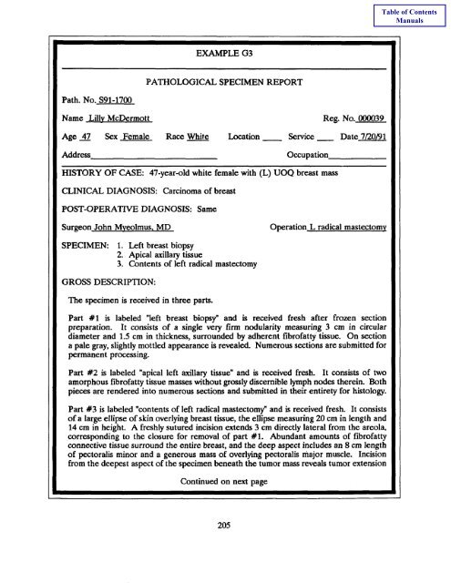
- Page 204 and 205: Example G1 can be abstracted as fol
- Page 206 and 207: Abstracting the Pathology Report Le
- Page 208 and 209: EXAMPLE G2 PATHOLOGY REPORT Path. N
- Page 212 and 213: EXAMPLE G4 DEPARTMENT OF PATHOLOGY
- Page 214 and 215: EXAMPLE (34 (continued) DIAGNOSIS:
- Page 216 and 217: EXAMPLE G5 PATHOLOGICAL SPECIMEN RE
- Page 218 and 219: The information which should be abs
- Page 220 and 221: PRACTICAL EXERCISE In the following
- Page 222 and 223: EXAMPLE G6 PATHOLOGY REPORT Name Be
- Page 224 and 225: EXAMPLE G7 PATHOLOGICAL SPECIMEN RE
- Page 226 and 227: EXAMPLE G8 DEPARTMENT OF PATHOLOGY
- Page 228 and 229: EXAMPLE G9 DEPARTMENT OF PATHOLOGY
- Page 230 and 231: PRACTICAL EXERCISE ANSWERS Example
- Page 232 and 233: EXAMPLE G10 AUTOPSY REPORT IDENTIFI
- Page 234 and 235: EXAMPLE G10 (continued) CLINICOPATH
- Page 236 and 237: Example G10 can be abstracted as fo
- Page 238 and 239: EXAMPLE Gll DEPARTMENT OF PATHOLOGY
- Page 240 and 241: EXAMPLE G11 (Continued) DEPARTMENT
- Page 242 and 243: EXAMPLE Gll (Continued) DEPARTMENT
- Page 244 and 245: Example G11 can be abstracted as fo
- Page 246 and 247: CYTOLOGIC EXAMINATION The study of
- Page 248 and 249: A cytology report recorded as suspi
- Page 250 and 251: PRACTICAL EXERCISE Summarize the fi
- Page 252 and 253: EXAMPLE G13 i ! PATIENT NO. DATE 10
- Page 254 and 255: EXAMPLE G14 Patient No. 000050 DATE
- Page 256 and 257: EXAMPLE GI_ CLINICAL RECORD - TISSU
- Page 258 and 259: Examples G12-15 may be abstracted a
- Page 260 and 261:
COMMON ABBREVIATIONS Abbreviation T
- Page 262 and 263:
HEENT Head, _ eara, ame & throat LE
- Page 264 and 265:
SARC Sarcoma UMB Navel (umbilicus)
- Page 266 and 267:
COMMON ABBREVIATIONS Definition Ind
- Page 268 and 269:
Impression IMP Lumbar spine L-SPINE
- Page 270 and 271:
Staphylococcus STAPH Within normal
- Page 272 and 273:
COMMON SYMBOLS Symbol Index Symbol
- Page 274 and 275:
ACRONYMS FOR ORGANIZATIONS CONCERNE
- Page 276 and 277:
WORLDWIDE ORGANIZATIONS IACR Intern
- Page 278 and 279:
ACRONYMS FOR STUDY GROUPS The follo
- Page 280 and 281:
SELECTED BIBLIOGRAPHY Cancer Regist
- Page 282 and 283:
INDEX Abstract, 3, 9, 11, 15-17, 21
- Page 284 and 285:
Cerebral angiogram, 40 Chemistry (S
- Page 286 and 287:
Endoscopic examination (Cont'd) Exa
- Page 288 and 289:
Laryngogram, 65, 69 Example E9, 65,
- Page 290 and 291:
Papanicolaou classification, 243 Pa
- Page 292 and 293:
Special examinations, 8 SPECT, 71 S
- Page 294 and 295:
Index of Examples E_mmple E1 29, 31