Self Instructional Manual for Cancer Registrars - SEER - National ...
Self Instructional Manual for Cancer Registrars - SEER - National ...
Self Instructional Manual for Cancer Registrars - SEER - National ...

Create successful ePaper yourself
Turn your PDF publications into a flip-book with our unique Google optimized e-Paper software.
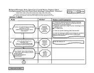
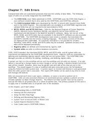
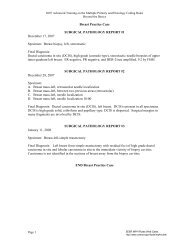
EXAMPLE<br />
G4<br />
DEPARTMENT<br />
OF PATHOLOGY<br />
Path. No. $91-0100<br />
Name Phineas Feltbad Reg. No. 000040<br />
Age 79 Sex Male Race White Location Service<br />
Date 5/23/91<br />
Address Occupation Drugllist<br />
PREOPERATIVE DIAGNOSIS: <strong>Cancer</strong> of left floor of mouth<br />
OPERATIVE FINDINGS: <strong>Cancer</strong> of left floor of mouth<br />
POSTOPERATIVE DIAGNOSIS: Same<br />
GROSS:<br />
The specimen<br />
SURGICAL PATHOLOGY SECTION<br />
is received in three parts, all fresh.<br />
Part #1 which is labeled "? metastatic tumor in jugular vein lymph node" consists of an<br />
elliptical fragment of light whitish-tan tissue which measures approximately 0.3 x 0.2 x 0.2<br />
cm. The specimen is examined by the frozen section technique, and the diagnosis is<br />
"ganglion." The remainder of part #1 of the specimen is submitted as frozen section control<br />
#1.<br />
Part #2 is labeled "resection of floor of mouth continuous with tongue and mandible plus<br />
left radical neck dissection." As received in the frozen section room, the specimen consists<br />
of a grossly identifiable left radical neck dissection and also the entire left ascending ramus<br />
of the mandible, the posterior three-fourths of the left mandible proper, the left lateral<br />
portion of the tongue, and the submental and submaxillary salivary glands. The main lesion<br />
is identified on the left side of the floor of the mouth. There is a crater<strong>for</strong>m lesion which<br />
measures approximately 1.2 x 0.5 cm in greatest dimensions. With the assistance of Dr. U.<br />
No Whoo, the specimen is properly oriented. Two areas of interest are defined. The first<br />
of these is the anterior tongue margin. The second of these is the medial tongue margin.<br />
Fragments from each of these areas are examined by the frozen section technique. The<br />
diagnosis on frozen section #2 (anterior tongue margin) is "no tumor seen" and on frozen<br />
section #3 (medial tongue margin) is "no tumor seen. N Two additional areas of special<br />
interest are identified. The first of these is that portion of the left radical neck dissection<br />
which was nearest to the carotid artery. A fragment of tissue from this area is excised and<br />
submitted <strong>for</strong> sectioning labeled "CM." The second area of interest is that portion of the<br />
Continued on next page<br />
207