Self Instructional Manual for Cancer Registrars - SEER - National ...
Self Instructional Manual for Cancer Registrars - SEER - National ...
Self Instructional Manual for Cancer Registrars - SEER - National ...

You also want an ePaper? Increase the reach of your titles
YUMPU automatically turns print PDFs into web optimized ePapers that Google loves.
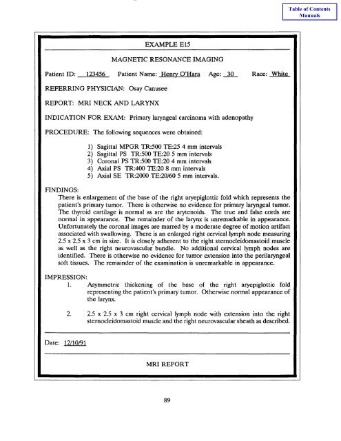
EXAMPLE<br />
El5<br />
MAGNETIC RESONANCE IMAGING<br />
Patient ID: 123456 Patient Name: Henry_ O'Hara Age: 30 Race: White<br />
REFERRING PHYSICIAN: Osay Canusee<br />
REPORT: MRI NECK AND LARYNX<br />
INDICATION FOR EXAM: Primary laryngeal carcinoma with adenopathy<br />
PROCEDURE: The following sequences were obtained:<br />
FINDINGS:<br />
1) Sagittal MPGR TR:500 TE:25 4 mm intervals<br />
2) Sagittal PS TR:500 TE:20 5 mm intervals<br />
3) Coronal PS TR:500 TE:20 4 mm intervals<br />
4) Axial PS TR:400 TE:20 8 mm intervals<br />
5) Axial SE TR:2000 TE:20/60 5 mm intervals.<br />
There is enlargement of the base of the right aryepiglottic fold which represents the<br />
patient's primary tumor. There is otherwise no evidence <strong>for</strong> primary laryngeal tumor.<br />
The thyroid cartilage is normal as are the arytenoids. The true and false cords are<br />
normal in appearance. The remainder of the larynx is unremarkable in appearance.<br />
Un<strong>for</strong>tunately the coronal images are marred by a moderate degree of motion artifact<br />
associated with swallowing. There is an enlarged right cervical lymph node measuring<br />
2.5 x 2.5 x 3 cm in size. It is closely adherent to the right sternocleidomastoid muscle<br />
as well as the right neurovascular bundle. No additional cervical lymph nodes are<br />
identified. There is otherwise no evidence <strong>for</strong> tumor extension into the perilaryngeal<br />
soft tissues. The remainder of the examination is unremarkable in appearance.<br />
IMPRESSION:<br />
1. Asymmetric thickening of the base of the right aryepiglottic fold<br />
representing the patient's primary tumor. Otherwise normal appearance of<br />
the larynx.<br />
2. 2.5 x 2.5 x 3 cm right cervical lymph node with extension into the right<br />
sternocleidomastoid muscle and the right neurovascular sheath as described.<br />
Date: 12/10/91<br />
MRI<br />
REPORT<br />
89