Self Instructional Manual for Cancer Registrars - SEER - National ...
Self Instructional Manual for Cancer Registrars - SEER - National ...
Self Instructional Manual for Cancer Registrars - SEER - National ...

Create successful ePaper yourself
Turn your PDF publications into a flip-book with our unique Google optimized e-Paper software.
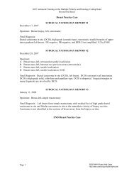
EXAMPLE G3 continued<br />
grossly to within 0.5 cm of muscle. Sections are submitted according to the following<br />
code: DE - deep surgical resection margins; SU, LA, INF, ME -- full thickness radial<br />
samplings from the center of the tumor superiorly, laterally, inferiorly and medially,<br />
respectively; NI - nipple and subjacent tissue. Lymph nodes dissected free from axillary<br />
fibrofatty tissue from levels I, II, and III will be labeled accordingly.<br />
MICROSCOPIC:<br />
Sections of part #1 confirm frozen section diagnosis of infiltrating duct carcinoma. It is<br />
to be noted that the tumor cells show considerable pleomorphism, and mitotic figures are<br />
frequent (as many as 4 per high power field). Many foci of calcification are present within<br />
the tumor.<br />
Part #2 consists of fibrofatty tissue and a single tiny lymph node free of disease.<br />
Part #3 includes 18 lymph nodes, three from Level III, two from Level II and thirteen<br />
from Level I. All lymph nodes are free of disease with the exception of one Level I<br />
lymph node which contains several masses of metastatic carcinoma.<br />
All sections taken radially from the superficial center of the resection site fail to include<br />
tumor, indicating the tumor to have originated deep within the breast parenchyma.<br />
Similarly, there is no malignancy in the nipple region, or in the lactiferous sinuses.<br />
Sections of deep surgical margin demonstrate diffuse tumor infiltration of deep fatty<br />
tissues, however, there is no invasion of muscle. Total size of primary tumor is estimated<br />
to be 4 cm in greatest dimension.<br />
DIAGNOSIS:<br />
1. Infiltrating duct carcinoma, left breast.<br />
2. Metastatic carcinoma, left axillary lymph node (1), Level I.<br />
3. Lymph nodes, no pathologic diagnosis, left axilla, Level I (12), Level II (2), Level III<br />
(3).<br />
Justin A. Glance, MD<br />
Pathologist<br />
Name Lilly McDermott Reg. No. 000039<br />
2O6