Self Instructional Manual for Cancer Registrars - SEER - National ...
Self Instructional Manual for Cancer Registrars - SEER - National ...
Self Instructional Manual for Cancer Registrars - SEER - National ...

Create successful ePaper yourself
Turn your PDF publications into a flip-book with our unique Google optimized e-Paper software.
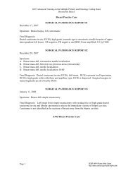
EXAMPLE G10 (continued)<br />
CLINICOPATHOLOGICAL CORRELATION AND DIAGNOSIS<br />
This 70-year-old white man was diagnosed in May 1982 at age 61 as having chronic<br />
lymphocytic leukemia. He received one intravenous injection of nitrogen mustard with good<br />
results.<br />
In January 1983, the patient was admitted to the hospital <strong>for</strong> evaluation and his white<br />
blood count was 5200 with 55% lymph., 37% mature polymorphs, 4% monocytes and 2%<br />
eosinophils. He was asymptomatic, and his prognosis <strong>for</strong> the future was described as<br />
excellent. In November 1991, the patient was admitted to the hospital, about 18 days be<strong>for</strong>e<br />
his death. There the diagnosis of undifferentiated adenocarcinoma of the stomach with<br />
massive dissemination of the disease was made. The patient died on December 1, 1991.<br />
Microscopic examination showed a signet ring cell carcinoma of stomach infiltrating the<br />
muscular coat with fibroblastic proliferation and thickening of the serosa. Metastatic signet<br />
ring cell carcinoma was found in "many organs (see final pathologic diagnosis) and<br />
represented massive widespread metastatic disease.<br />
Only microscopically were we able to diagnose adenocarcinoma of the right bronchus,<br />
locally invasive and infiltrating bronchial cartilage and adjacent nerves but without<br />
metastases.<br />
Although, the patient was free of any symptoms of chronic lymphocytic leukemia, sections<br />
of the liver and bone marrow showed lymphocytic leukemic infiltrates. The enlargement of<br />
the spleen (770 gm.) and lymph nodes might be due to metastatic signet ring cell carcinoma<br />
as well as to leukemic involvement.<br />
At autopsy this case revealed three histologically different tumors. Two of them were<br />
carcinomas: signet ring cell carcinoma of the stomach with widespread metastases and<br />
adenocarcinoma of the right bronchus with local infiltration. The third malignancy was<br />
chronic lymphocytic leukemic infiltrates without definite clinical manifestation. It is quite<br />
possible, that the signet ring cell carcinoma with metastases, diagnosed in late stage of such<br />
disease, covered other slightly marked features of leukemia or adenocareinoma of the lung.<br />
The myelofibrosis could be secondary to metastatic signet ring cell carcinoma or the results<br />
of the previous leukemic chemotherapy or connected with leukemic involvement of the<br />
bone marrow, or all of them caused such condition.<br />
Doe, John Hosp. No. 000046 Autopsy #A91-21<br />
229