XVIII Congress of the Italian Society for Hemostasis and Thrombosis Research, Rome, Sept. 30-Oct. 3, <strong>2004</strong>21CO-0042-SUBSTITUTED ADENINE NUCLEOTIDES: MODULATION OFPLATELET AGGREGATION THROUGH P2Y RECEPTORSPodda GM, 1 Vittori S, Lecchi A, 1 Costanzi S, 2Cristalli G, 2 Cattaneo M 31Centro Emofilia e Trombosi Angelo Bianchi Bo<strong>no</strong>miIRCCS Ospedale Maggiore, Departimento diMedicina Interna, Università di Mila<strong>no</strong>, 2 Dipartimentodi Scienze Chimiche, Università di Cameri<strong>no</strong>;3Unità di Ematologia e Trombosi, Ospedale SanPaolo, Departimento di Medicina, Chirurgia eOdontoiatria, Università di Mila<strong>no</strong>, ItalyAim of our study was to evaluate the response ofhuman platelets to nucleotide analogues, in thepresence or absence of ADP. We tested the six compoundsreported in Figure 1. The compounds (mo<strong>no</strong>–,di– and tri-phosphate derivatives of 2-hexynyl and2-phenylethynylade<strong>no</strong>sine) were synthetizedthrough phosphorylation of the corresponding nucleosides.Shape change, aggregation and inhibition ofadenylyl cyclase induced by 10 microM ADP werestudied in washed human platelet suspensions.Nucleotides 2 and 3 caused platelet shape changeand aggregation, with an EC50 ranging from 10 to100 microM. The extent of platelet aggregationinduced by 100 microM of these compounds wasslightly lower than that elicited by 10 microM ADP,indicating that they activate both P2Y1 and P2Y12receptors. The 2-phenylethynylade<strong>no</strong>sine derivatives4-6 and the 2-hexynyl mo<strong>no</strong>phosphate derivative 1,at concentrations between 10 and 130 µM, did <strong>no</strong>tinduce platelet shape change or aggregation. In contrast,they concentration-dependently inhibitedplatelet aggregation induced by 10 µM ADP, withthe following IC(sub)50: 14 µM (1), 130 µM (4), 60µM (5) and 85 µM (6). At a concentration of 800microM, nucleotides 1, 4 and 6 completely inhibitedboth platelet aggregation and shape change inducedby 10 µM ADP, while nucleotide 5 inhibited plateletaggregation by 90% without affecting shape change.At a concentration of 800 µM, all the nucleotidespartly (30-75%) antagonized the ADP-induced inhibitio<strong>no</strong>f cAMP increase; lower concentrations had<strong>no</strong> inhibitory effects. Therefore the tested inhibitorynucleotides displayed primarily an antagonisticeffect on the platelet P2Y1 receptor, while a partialinhibitory effect on the platelet P2Y12 receptor wasobserved only at the highest (800 µM) nucleotideconcentrations tested. The surprisingly differentbehaviour of 2, 3 (agonists) and 5, 6 (antagonists)can be explained in terms of different size and flexibilityof the substituent in 2 position.CO-005ROLE OF THE PLATELET P2Y1 RECEPTOR IN THROMBUSFORMATION UNDER FLOW CONDITIONSMarchese P,* Cattaneo M, # Gachet C,^ Ruggeri ZM**Division of Experimental Thrombosis andHemostasis, The Scripps Research Institute, La Jolla,California, USA; # Unit of Hematology and Thrombosis,Department of Surgery, Medicine and Dentistry,Ospedale San Paolo, University of Milan, Italy;^Institut National de la Sante et de la RechercheMedicale, Etablissement Francais du Sang-Alsace,Strasbourg, FranceP2Y1 is a receptor for ade<strong>no</strong>sine diphosphate (ADP)on platelets required for <strong>no</strong>rmal shape change andaggregation following ADP stimulation. Moreover,P2Y1 deficient mice display defective platelet aggregationin response to ADP or low collagen concentrations,as well as reduced thrombus formation inarterial and ve<strong>no</strong>us models of vascular injury. Tocharacterize in more detail the role of P2Y1 inplatelet function, we tested P2Y1 deficient mouseblood in an ex vivo model of thrombus formation onbovine insoluble fibrillar collagen type I under flowconditions. In the absence of P2Y1, platelets formedsmaller than <strong>no</strong>rmal thrombi on surfaces with eitherlow or high collagen density when the wall shearrate was 1500 s -1 , but the defect was <strong>no</strong>t detectableat the lower shear rates of 300 s -1 and 500 s(e) 1. Athigher shear rates, 6000-10000 s -1 , the defect waspresent at low but less apparent at higher collagenconcentrations. Real-time analysis of thrombus formationdemonstrated that the initial platelet adhesionand aggregation, within 30 s from the beginningof perfusion, were <strong>no</strong>rmal or only partially reduced,respectively, in the absence of P2Y1, while thereduced thrombus volume as compared to <strong>no</strong>rmalbecame progressively more apparent at later perfusiontimes. In fact, defective thrombus formation inthe absence of P2Y1, when present, was characterizedby instability of initially formed thrombi thattended to detach from the surface. Thus, the P2Y1ADP receptor plays a role in collagen-induced thrombusformation at medium to high shear rates by substantiallycontributing to thrombus stabilization.These results also indicate that P2Y1 function maybe more relevant for <strong>no</strong>rmal platelet function atmedium to high, rather than low, wall shear rates,and particularly on surfaces with lower collagen density.haematologica vol. <strong>89</strong>(suppl. n. 8):september <strong>2004</strong>
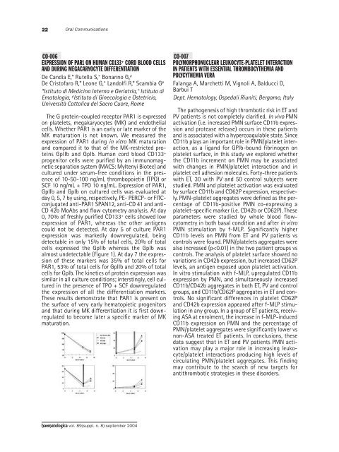
22Oral CommunicationsCO-006EXPRESSION OF PAR1 ON HUMAN CD133 + CORD BLOOD CELLSAND DURING MEGACARYOCYTE DIFFERENTIATIONDe Candia E,* Rutella S,° Bonan<strong>no</strong> G, #De Cristofaro R,* Leone G,° Landolfi R,* Scambia G #*Istituto di Medicina Interna e Geriatria,° Istituto diEmatologia, # Istituto di Ginecologia e Ostetricia,Università Cattolica del Sacro Cuore, RomeThe G protein-coupled receptor PAR1 is expressedon platelets, megakaryocytes (MK) and endothelialcells. Whether PAR1 is an early or late marker of theMK maturation is <strong>no</strong>t k<strong>no</strong>wn. We measured theexpression of PAR1 during in vitro MK maturationand compared it to that of the MK-restricted proteinsGpIIb and GpIb. Human cord blood CD133 +progenitor cells were purified by an immu<strong>no</strong>magneticseparation system (MACS: Myltenyi Biotec) andcultured under serum-free conditions in the presenceof 10-50-100 ng/mL thrombopoietin (TPO) orSCF 10 ng/mL + TPO 10 ng/mL. Expression of PAR1,GpIIb and GpIb on cultured cells was evaluated atday 0, 5, 7 by using, respectively, PE- PERCP- or FITCconjugatedanti-PAR1 SPAN12, anti-CD 41 and anti-CD 42b MoAbs and flow cytometry analysis. At day0, 70% of freshly purified CD133 + cells showed lowexpression of PAR1, whereas the other antigenscould <strong>no</strong>t be detected. At day 5 of culture PAR1expression was markedly downregulated, beingdetectable in only 15% of total cells, 20% of totalcells expressed the GpIIb whereas the GpIb wasalmost undetectable (Figure 1). At day 7 the expressio<strong>no</strong>f these markers was 35% of total cells forPAR1, 53% of total cells for GpIIb and 20% of totalcells for GpIb. The kinetics of protein expression wassimilar in all culture conditions; interstingly, cell culturedin the presence of TPO + SCF downregulatedthe expression of all the differentiation markers.These results demonstrate that PAR1 is present onthe surface of very early hematopietic progenitorsand that during MK differentiation it is first downregulatedto become later a specific marker of MKmaturation.CO-007POLYMORPHONUCLEAR LEUKOCYTE-PLATELET INTERACTIONIN PATIENTS WITH ESSENTIAL THROMBOCYTHEMIA ANDPOLYCYTHEMIA VERAFalanga A, Marchetti M, Vig<strong>no</strong>li A, Balducci D,Barbui TDept. Hematology, Ospedali Riuniti, Bergamo, ItalyThe pathogenesis of high thrombotic risk in ET andPV patients is <strong>no</strong>t completely clarified. In vivo PMNactivation (i.e. increased PMN surface CD11b expressionand protease release) occurs in these patientsand is associated with a hypercoagulable state. SinceCD11b plays an important role in PMN/platelet interaction,as a ligand for GPIb-bound fibri<strong>no</strong>gen onplatelet surface, in this study we explored whetherthe CD11b increment on PMN may be associatedwith changes in PMN/platelet interaction and inplatelet cell adhesion molecules. Forty-three patientswith ET, 30 with PV and 50 control subjects werestudied. PMN and platelet activation was evaluatedby surface CD11b and CD62P expression, respectively.PMN-platelet aggregates were defined as the percentageof CD11b-positive PMN co-expressing aplatelet-specific marker (i.e. CD42b or CD62P). Theseparameters were studied by whole blood flowcytometryin both basal condition and after in vitroPMN stimulation by f-MLP. Significantly higherCD11b levels on PMN from ET and PV patients vscontrols were found. PMN/platelets aggregates werealso increased (p