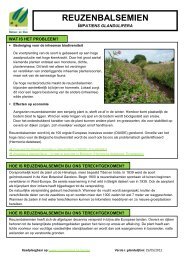
LABOULBENIALES - Agentschap voor Natuur en Bos
LABOULBENIALES - Agentschap voor Natuur en Bos
LABOULBENIALES - Agentschap voor Natuur en Bos

Create successful ePaper yourself
Turn your PDF publications into a flip-book with our unique Google optimized e-Paper software.
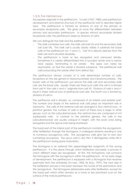
1.3.3. THE PERITHECI UMAscospores originate in the perithecium. TAVARES (1967, 1985) used peritheciumdevelopm<strong>en</strong>t and external structure of the perithecial wall to describe highertaxa. The perithecium is formed by divisions of one of the primary orsecondary receptacle cells. This gives at once the differ<strong>en</strong>tiation betwe<strong>en</strong>primary and secondary perithecium. In species without secondarily dividedreceptacle cells, the perithecium arises by divisions of cell II.We can distinguish the stalk and the perithecium:The stalk comprises two cells, the stalk cell (cell VI) and the secondary stalkcell (cell VII). The stalk cell is usually clearly visible; it subt<strong>en</strong>ds the basalcells of the perithecium (m, n and n’). Cell VII is directly derived from thestalk cell and is situated abaxially.The perithecium is more or less elongated and narrowed distally.Sometimes it is clearly differ<strong>en</strong>tiated into a rounded v<strong>en</strong>ter and a narrowneck (apex), terminating in an ostiole. The apex can rarely beasymmetric, so that the ostiole is situated subapical. The perithecial wallcells surrounding the ostiole oft<strong>en</strong> form distinct lips.The perithecium always consists of a well determined number of cells.Exceptions on this are g<strong>en</strong>era in Herpomycetidae and Ceratomycetidae. Thelowest cells of the perithecium, just above the stalk cell and secondary stalkcell, are the basal cells. Usually, there are three such cells: the cell m is formedfrom cell VI, the cells n and n’ originate from cell VII. Divisions of cells n and n’result in three vertical rows of perithecial wall cells. The fourth row is formed bydivisions of cell m.The perithecial wall is double, i.e. composed of an interior and exterior part.The number and shape of the external wall cells plays an important role intaxonomy. The cells of the external wall are arranged in four vertical rows. Inprimitive g<strong>en</strong>era, the number of cells in each of these rows is large; in othergroups, such as the Laboulb<strong>en</strong>iaceae, the vertical rows are composed of 4-5superposed cells. In contrast to the primitive g<strong>en</strong>era, the cells in theLaboulb<strong>en</strong>iaceae are usually unequal in height, with the lower ones beingelongated and the apical ones being short<strong>en</strong>ed.The basal part of the interior part of the perithecium contains the ascogonium.After fertilization through the trichogyne, it undergoes divisions resulting in oneto numerous ascog<strong>en</strong>ous cells. The ascog<strong>en</strong>ous cells give rise to new ascicontaining ascospores. The ascus wall is very thin; it disintegrates while still inthe perithecium during spore maturation.The trichogyne is an external thin app<strong>en</strong>dage-like outgrowth of the youngperithecium. It is the place through which fertilization proceeds, a process inwhich differ<strong>en</strong>t steps are recognized. At first, the trichophoric cell connectsthe trichogyne with the ascogonium within the perithecium. At an early stageof developm<strong>en</strong>t, the perithecium is equipped with a trichogyne that receivesspermatia from the antheridia (TAVARES, 1985; DE KESEL, 1997). The next step inthe fertilization process is the fusion betwe<strong>en</strong> the nuclei of the spermatium andthe ascogonium. The trichogyne deteriorates early after fertilization, except forthe basal part which oft<strong>en</strong> remains as a more or less promin<strong>en</strong>t scar on thesurface of the mature perithecium.P a g e | 13