Tumorile cardiopericardice Prof Dr Grigore Tinica, Dr. Lutea Mirela ...
Tumorile cardiopericardice Prof Dr Grigore Tinica, Dr. Lutea Mirela ...
Tumorile cardiopericardice Prof Dr Grigore Tinica, Dr. Lutea Mirela ...

You also want an ePaper? Increase the reach of your titles
YUMPU automatically turns print PDFs into web optimized ePapers that Google loves.
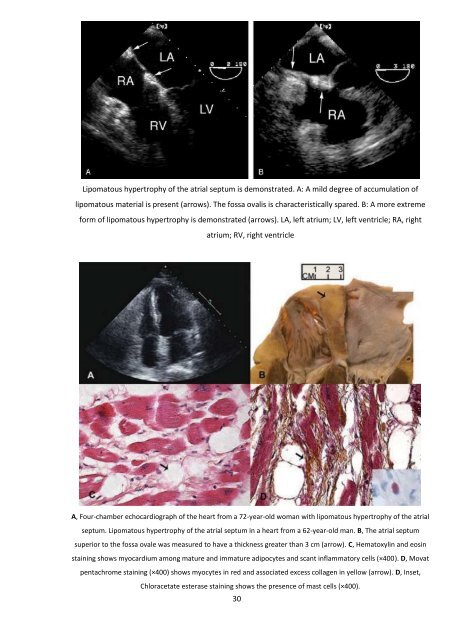
Lipomatous hypertrophy of the atrial septum is demonstrated. A: A mild degree of accumulation of<br />
lipomatous material is present (arrows). The fossa ovalis is characteristically spared. B: A more extreme<br />
form of lipomatous hypertrophy is demonstrated (arrows). LA, left atrium; LV, left ventricle; RA, right<br />
atrium; RV, right ventricle<br />
A, Four-chamber echocardiograph of the heart from a 72-year-old woman with lipomatous hypertrophy of the atrial<br />
septum. Lipomatous hypertrophy of the atrial septum in a heart from a 62-year-old man. B, The atrial septum<br />
superior to the fossa ovale was measured to have a thickness greater than 3 cm (arrow). C, Hematoxylin and eosin<br />
staining shows myocardium among mature and immature adipocytes and scant inflammatory cells (×400). D, Movat<br />
pentachrome staining (×400) shows myocytes in red and associated excess collagen in yellow (arrow). D, Inset,<br />
Chloracetate esterase staining shows the presence of mast cells (×400).<br />
30