medtropoleAktuelles aus der Klinik für einweisende Ärzte - Asklepios
medtropoleAktuelles aus der Klinik für einweisende Ärzte - Asklepios
medtropoleAktuelles aus der Klinik für einweisende Ärzte - Asklepios

Sie wollen auch ein ePaper? Erhöhen Sie die Reichweite Ihrer Titel.
YUMPU macht aus Druck-PDFs automatisch weboptimierte ePaper, die Google liebt.
a<br />
Literatur<br />
[1] Saqqur M, Uchino K, Demchuk AM, et al. Site of arterial<br />
occlusion identified by transcranial Doppler predicts the<br />
response to intravenous thrombolysis for stroke. Stroke<br />
2007; 38(3): 948-54.<br />
[2] Stolz E, Cioli F, Allendoerfer J, Gerriets T, Del Sette M,<br />
Kaps M. Can early neurosonology predict outcome in<br />
acute stroke? A metaanalysis of prognostic clinical effect<br />
sizes related to the vascular status. Stroke 2008; 39: 3255-<br />
61.<br />
[3] Warfarin-Aspirin Symptomatic Intracranial Disease<br />
(WASID) Trial Investigators. Design, progress and challenges<br />
of a double-blind trial of warfarin versus aspirin for<br />
symptomatic intracranial arterial stenosis. Neuroepidemiology<br />
2003; 22: 106-17.<br />
[4] Seidel G, Kaps M, Gerriets T. Potential and limitations<br />
of transcranial color-coded sonography in stroke patients.<br />
Stroke 1995; 26: 2061-6.<br />
[5] Mäurer M, Shambal S, Berg D, et al. Differentiation<br />
between intracerebral hemorrhage and ischemic stroke by<br />
transcranial color-coded duplex-sonography. Stroke 1998;<br />
29: 2563-7.<br />
b<br />
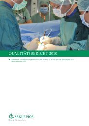
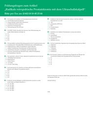
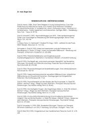
Abb. 4<br />
a: CCT-Kontrolle am 2. Erkrankungstag mit demarkiertem insulären Hirninfarkt (roter Pfeil) bei einem Patienten<br />
mit akutem Mediaverschluss. Microvascular Imaging (MVI) zwei Stunden nach Symptombeginn mit signalintenser<br />
Darstellung des durchbluteten Hirngewebes (b). Das infarzierte Hirngewebe ist nicht von Kontrastmittel durchflossen<br />
und damit schwarz dargestellt. Zu diesem Zeitpunkt zeigte das initiale CCT noch keine Auffälligkeiten. Die gelbe<br />
Markierung zeigt die Beschallungsebene des MVI.<br />
[6] Seidel G, Cangür H, Albers T, Burgemeister A, Meyer-<br />
Wiethe K. Sonographic evaluation of hemorrhagic transformation<br />
and arterial recanalization in acute hemispheric<br />
ischemic stroke. Stroke 2009; 40: 119-23.<br />
[7] Gerriets T, Stolz E, König S, et al. Sonographic monitoring<br />
of midline shift in space-occupying stroke - an early<br />
outcome predictor. Stroke 2001; 32: 442-7.<br />
[8] Seidel G, Meyer-Wiethe K, Berdien G, Hollstein D,<br />
Toth D, Aach T. Ultrasound perfusion imaging in acute<br />
middle cerebral artery infarction predicts outcome. Stroke.<br />
2004; 35: 1107-11.<br />
[9] Eggers J, König IR, Koch B, Händler G, Seidel G.<br />
Sonothrombolysis with transcranial color-coded sonography<br />
and recombinant tissue-type plasminogen activator in<br />
acute middle cerebral artery main stem occlusion. Results<br />
from a randomized study. Stroke 2008; 39: 1470-5.<br />
[10] Eggers J, Seidel G, Koch B, König IR. Sonothrombolysis<br />
in acute ischemic stroke for patients ineligible for rt-PA.<br />
Neurology 2005; 64: 1052-4.<br />
Kontakt<br />
Prof. Dr. Günter Seidel<br />
Abteilung Neurologie<br />
<strong>Asklepios</strong> <strong>Klinik</strong> Nord<br />
Tangstedter Landstraße 400<br />
22417 Hamburg<br />
Tel. (0 40) 18 18-87 30 76<br />
Fax (0 40) 18 18-87 30 69<br />
E-Mail: g.seidel@asklepios.com<br />
Neurologie<br />
911