

Comparative Parasitology 67(1) 2000 - Peru State College
Comparative Parasitology 67(1) 2000 - Peru State College
Comparative Parasitology 67(1) 2000 - Peru State College

Create successful ePaper yourself
Turn your PDF publications into a flip-book with our unique Google optimized e-Paper software.
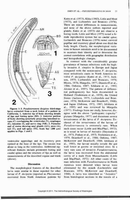
Figures 1-3. Pseudoterranova decipiens third-stage<br />
larva extracted from a neck lesion of a patient in<br />
California. 1. Anterior tip of body showing details<br />
of lips and boring spine (BS). 2. Anterior portion<br />
of body showing anteriorly projecting intestinal cecum<br />
(C) overlapping the ventriculus (V), esophagus<br />
(E), intestine (I), and nerve ring (NR). 3. Posterior<br />
end of worm showing anal glands (AG), anus (A),<br />
tail (T), and tail spine (TS). Scale bar (200 u-m)<br />
applies to Figs. 1 and 3.<br />
ventricular appendage and its excretory pore<br />
opened at the base of the lips. The cecum was<br />
about as long as the ventriculus. Additional significant<br />
features include prominent boring and<br />
caudal spines and the structure of lips as well as<br />
measurements of the described organs and trunk<br />
(above).<br />
Discussion<br />
The morphological features of the described<br />
larva were similar to those reported for other<br />
larvae of P. decipiens (reported as Phocanemd)<br />
recovered from North American patients by<br />
AMIN ET AL.—PSEUDOTERRANOVA IN MAN 73<br />
Kates et al. (1973), Kliks (1983), Little and Most<br />
(1973), and Lichtenfels and Brancato (1976).<br />
There are minor differences in measurements,<br />
and none of the above authors reported anal<br />
glands; Kates et al. (1973) did not observe a<br />
boring tooth; Little and Most (1973) noted a female<br />
reproductive system but no caudal spine;<br />
Lichtenfels and Brancato (1976) noted cervical<br />
papillae and excretory gland about one-third the<br />
body length. Clearly, the morphological variations<br />
in human anisakids need to be documented<br />
to ascertain their identity and to determine the<br />
correct correlations with geographic distribution<br />
and histopathologic changes.<br />
In contrast with the considerably greater<br />
prevalence of human infections with the highly<br />
invasive A. simplex in Europe and Japan<br />
compared with the noninvasive P. decipiens,<br />
most anisakiasis cases in North America involve<br />
P. decipiens (Kates et al., 1973; Jackson,<br />
1975; Lichtenfels and Brancato, 1976;<br />
Myers, 1976; Margolis, 1977; Deardorff et al.,<br />
1986; Oshima, 1987; Ishikura et al., 1993;<br />
Alonso et al., 1997). The pattern of differential<br />
pathogenicity has been documented in<br />
Holland (Yoshimura et al., 1979), the United<br />
<strong>State</strong>s (Jackson, 1975; Lichtenfels and Brancato,<br />
1976; McKerrow and Deardorff, 1988),<br />
and Japan (Oshima, 1972, 1987; Ishikura et<br />
al., 1993) and was reviewed by Margolis<br />
(1977). Findings from our study, however, disagree<br />
with the above "pathogenic capacity"<br />
picture (Margolis, 1977) and document severe<br />
invasiveness of the larva of P. decipiens. Evidence<br />
of the invasiveness of the larvae of<br />
Pseudoterranova is extremely rare. When<br />
such cases occur (Little and MacPhail, 1972),<br />
as is usual in larvae of Anisakis (Hayasaka et<br />
al., 1971; Pinkus et al., 1975; Yoshimura et al.,<br />
1979; Deardorff et al., 1986; Oshima, 1987;<br />
McKerrow and Deardorff, 1988; Ishikura et<br />
al., 1993), the larvae usually invade the gut<br />
wall lower at gastric or intestinal sites. In a<br />
very rare case of invasive Pseudoterranova,<br />
the larva was recovered from the abdominal<br />
cavity of a male from Massachusetts (Little<br />
and MacPhail, 1972). All other cases of human<br />
infection with Pseudoterranova in North<br />
America were detected when larvae were<br />
eliminated by the mouth (Lichtenfels and<br />
Brancato, 1976; McKerrow and Deardorff,<br />
1988). A larva was identified as "Anisakis"<br />
from histological sections in the tonsils of a<br />
Copyright © 2011, The Helminthological Society of Washington


