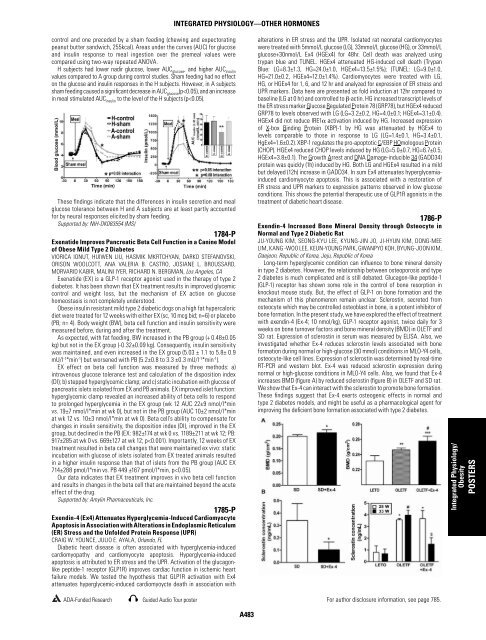
control and one preceded by a sham feeding (chewing and expectorating peanut butter sandwich, 255kcal). Areas under the curves (AUC) for glucose and insulin response to meal ingestion over the premeal values were compared using two-way repeated ANOVA. H subjects had lower nadir glucose, lower AUC glucose , and higher AUC insulin values compared to A group during control studies. Sham feeding had no effect on the glucose and insulin responses in the H subjects. However, in A subjects sham feeding caused a signifi cant decrease in AUC glucose (p
Integrated Physiology/ Obesity POSTERS 1787-P Implication of Corticotropin-Releasing-Hormone on INS-1 and Pancreatic Islets In Vitro BARBARA LUDWIG, JANINE SCHMID, CHRISTIAN ZIEGLER, MONIKA EHRHART- BORNSTEIN, STEFAN R. BORNSTEIN, Dresden, Germany Hormonal factors, including corticotropin-releasing-hormone (CRH) and glucocorticoids (GC) regulate the activity of the hypothalamic-pituitaryadrenal (HPA) axis in response to stress. This axis is kept in balance by the negative feedback effects of GC on the CRH synthesis and secretion in the hypothalamus. Recently CRH has been identifi ed to promote β-cell proliferation and potentiates insulin secretion in a glucose dependent manner. On the other hand, GCs are referred to as diabetogenic hormones due to the induction of gluconeogenesis, implication on development of insulin resistance, effects on adipocytes and the functionally insulinantagonizing effects. Therefore, an imbalance of the CRH/GC system may lead to metabolic dysfunction and diabetes. GC access to intracellular receptors is regulated by two isoforms of 11β-hydroxysteroid dehydrogenase (11β-HSD) which catalyse the interconversion of physiologically active GC to its inactive metabolite (11β-HSD-2) and vice versa (11β-HSD-1). In the present study we analyzed the mechanism of CRH mediated β-cell regulation and asked if pancreatic islets respond directly to CRH by interference with 11β-HSD activity and therefore GC activity. In in vitro studies we could demonstrate that CRH and its receptor are expressed on mRNA and protein levels in INS-1 cells, rat and human islets. We also found expression of both 11β-HSD isoforms in rat and human islets. CRH exposure of islets signifi cantly decreased mRNA levels of both 11β-HSD-isoforms as measured by quantitative RT-PCR. The specifi c iso-enzyme activity was analyzed by measuring the production of active GC. Following CRH treatment, active GC levels were signifi cantly reduced indicating an overbalance in favour of 11β-HSD-2 activity. Stimulation with CRH resulted in signifi cantly increased insulin secretion. Moreover, CRHreceptor activation caused a signifi cant increase of cell proliferation and reduced cell apoptosis. We suggest that CRH may not only be of signifi cance within the endocrine stress system for triggering and sustaining obesity and metabolic dysfunction, but may also play a direct role for glucose homoeostasis and the regulation of β-cell mass. 1788-P Improved Glycemic Control Enhances the Incretin Effect in Patients with Type 2 <strong>Diabetes</strong> ZHIBO AN, SADIA ALI, COLLEEN ROGGE, FAY HAILES, CATHY BAILEY, BRENDA WENSTRUP, BRIANNE REEDY, MARZIEH SALEHI, DAVID A. D’ALESSIO, Cincinnati, OH The incretin effect is impaired in type 2 diabetes, and diabetic subjects have diminished responses to incretins. However, it is not clear whether the defects are specifi c for incretin stimulated insulin secretion or simply another aspect of generalized β-cell dysfunction. Correction of chronic hyperglycemia improves incretin action in animals. Further, normalization of hyperglycemia in diabetic patients improves the potentiation of glucose-stimulated insulin secretion by GLP-1 and GIP. The aim of this study was to determine whether glycemic control specifi cally improves the incretin effect in humans. Six type 2 diabetic subjects with moderate to poor control (age 55±3; BMI 34±2) were studied twice using a glucose clamp-OGTT protocol before and after 8 weeks of longacting insulin treatment titrated to a fasting glucose target of 6.0 mM, causing Hg A1C to reduce from 8.2±0.2 to 6.8±0.2 %. Following an overnight fast and basal blood draws (-20-0 min), glucose was given intravenously (IV) to raise blood glucose by ∼6.5 mM. At 90 min 75 g of glucose was given orally, and the IV glucose infusion was adjusted to maintain the blood glucose constant. The incretin effect was calculated by comparing plasma insulin levels before and after the oral glucose load. In the basal period, P1 (0-90 min) and P2 (90- 270 min), the blood glucose levels were 8.6±0.6, 15.1±0.8, and 16.3±1.3 mM at visit 1; and 5.9±0.6, 13.2±0.8, and 14.3±1.0 mM at visit 2, respectively. The incremental insulin response to IV glucose (P1) increased from 0.10±0.06 (visit 1) to 0.16±0.08 nM (visit 2). The response to oral glucose ingestion at fi xed hyperglycemia (P2) increased from 0.42±0.30 (visit 1) to 0.90±0.38 nM (visit 2). After 8 weeks of treatment, the average increase of the incremental insulin response to oral glucose was 8 fold greater than the increase to IV glucose (p=0.2), with 5 out of 6 subjects having a 4 fold or greater increase. Our data support the hypothesis that intensifi ed insulin treatment to improve glycemic control leads to a disproportionate improvement of insulin secretion in response to oral, compared to isoglycemic IV, glucose stimulation in patients with type 2 diabetes. Supported by: VA Merit Award to D.D. For author disclosure information, see page 785. INTEGRATED PHYSIOLOGY—OTHER CATEGORY HORMONES A484 1789-P Involvement of Hypothalamic AMPK and Histamine on Olanzapine- Induced Glucose Intolerance in Mice MEGUMI ASATO, YOKO ISHIKAWA, HIROKO IKEDA, ATSUKO KAMEI, KENJI ONODERA, JUNZO KAMEI, Tokyo, Japan, Kanagawa, Japan Atypical antipsychotic drugs are well known to produce metabolic disturbance. Treatment with some atypical antipsychotic drugs such as clozapine or olanzapine induces the impairment of glucose metabolism, which increases the risk for developing metabolic side-effects, including weight gain, dyslipidemia, insulin resistance and hyperglycemia. We have already reported that central administration of olanzapine produces glucose intolerance. Recent study showed that clozapine activated adenosine 5’-monophosphate-activated protein kinase (AMPK) through histamine H1 receptors in the hypothalamus. AMPK is known to sense the energy status of cells and to regulate fuel availability. In central nervous system, AMPK modulates feeding and systemic energy homeostasis. Thus, it is possible that atypical antipsychotic drugs such as clozapine and olanzapine induces glucose intolerance by activating AMPK in the hypothalamus. Therefore, the present study was designed to investigate the involvement of histamine and AMPK on olanzapine-induced glucose intolerance. In the glucose CII test, both intraperitoneally (i.p.) or intracerebroventricually (i.c.v.) administration of olanzapine dose-dependently induced glucose intolerance as compared with the vehicle-treated group. The glucose intolerance induced by i.c.v. treatment with olanzapine was attenuated by i.p. pretreatment with histamine synthesis inhibitor, α-fl uoromethylhistidine (FMH). The glucose intolerance induced by i.c.v. treatment with olanzapine was also attenuated by i.c.v. pretreatment with an AMPK inhibitor, compound C. In addition, i.c.v. treatment with an AMPK activator, 5-aminoimidazole-4-carboxamide ribonucleoside (AICAR) also produced the same changes in serum glucose levels as olanzapine. In the western blot test, olanzapine increased the phosphorylation of AMPK in the hypothalamus, though total amount of AMPK was not affected. These results indicate that both histamine and AMPK in the hypothalamus are involved in the olanzapine-induced glucose intolerance. Furthermore, the present study suggests that olanzapine might activate hypothalamic AMPK via histaminergic system. 1790-P Low Estradiol Concentrations in Males with Hypogonadotrophic Hypogonadism and Type 2 <strong>Diabetes</strong> SANDEEP DHINDSA, RICHARD FURLANETTO, MEHUL VORA, HUSAM GHANIM, PARESH DANDONA, Buffalo, NY, Chantilly, VA One-third of men with type 2 diabetes have hypogonadotrophic hypogonadism(HH). It has been suggested that HH in these men may be due to an increase in plasma estradiol(E 2 ) concentrations secondary to an increase in aromatase activity in the adipose tissue which leads to the suppression of hypothalamo-hypophyseal-gonadal(HHG) axis. We investigated the hypothesis that plasma E 2 concentrations are signifi cantly greater in type 2 diabetic males with HH as compared to those without HH. Plasma estradiol, testosterone(T), LH and sex hormone binding globulin(SHBG) concentrations were measured in fasting blood samples of 236 men with type 2 diabetes (mean age: 56±12; range:23-83 years; mean BMI: 35±7;range:17-59kg/ m 2 ) attending a tertiary diabetes referral center. Total T was measured by liquid chromatography tandem mass spectrometry(LC-MS/MS). In 196 men, total E 2 was measured by an immunoassay. Free E 2 and T concentrations were calculated using total E 2 , T, albumin and SHBG. In 99 men, total E 2 was measured by the more specifi c LC-MS/MS assay. Free E 2 and free T concentrations in these men were measured by tracer equilibrium dialysis. HH was defi ned as free T