ECHINODERMATA - KU ScholarWorks - University of Kansas
ECHINODERMATA - KU ScholarWorks - University of Kansas
ECHINODERMATA - KU ScholarWorks - University of Kansas

You also want an ePaper? Increase the reach of your titles
YUMPU automatically turns print PDFs into web optimized ePapers that Google loves.
76 THE UNIVERSITY OF KANSAS PALEONTOLOGICAL CONTRIBUTIONS<br />
gins. Radials five, each hexagonal in side view, 5 mm.<br />
long by 3.5 mm. wide, with shallow sinus 4 mm. long by<br />
1.5 mm. wide, with growth striae subparallel to margins.<br />
Deltoids overlap radials.<br />
Deltoids four, lancet-shaped, each 3 mm. long by 2 mm.<br />
wide, notched at the adorai tip by two spiracles along<br />
ambulacral margins. On the anal side there are ?two<br />
deltoids; a horseshoe-shaped or V-shaped ?epideltoid<br />
adjacent to the oral opening, curving around the adorai<br />
part <strong>of</strong> the anal opening on three sides, and a large<br />
lancet-shaped hypodeltoid on the aboral side <strong>of</strong> the anal<br />
opening, extended ventrally into a hood or coronal process.<br />
Two cryptodeltoids may be present. The anal<br />
spiracles or hydrospire canals are separated internally<br />
from the anal opening by deltoid septa, but externally<br />
only one anispiracle is visible. Thus there are nine openings<br />
around the oral opening, consisting <strong>of</strong> eight spiracles<br />
and the anispiracle. There are three hydrospire folds on<br />
each side <strong>of</strong> an ambulacrum.<br />
Ambulacra five, broadly linear, each 7.5 mm. long<br />
by 1.5 mm. wide, each lancet plate being exposed onethird<br />
or more <strong>of</strong> its width along the middle <strong>of</strong> each<br />
ambulacrum. The side plates are normally disposed, with<br />
30 side plates in a space <strong>of</strong> 10 mm. along an ambulacrum,<br />
one pore between adjacent side plates notched in the<br />
deltoid and radial ambulacral margins, and three main<br />
cover-plate sockets to each side plate along the main food<br />
groove. In addition to fine growth striae, the calyx plates<br />
are ornamented by coarse ridges subparallel to the plate<br />
margins.<br />
Remarks.—The type specimens are grouped together<br />
in two glass vials and it has not been possible to find all<br />
<strong>of</strong> the figured specimens. There are 37 specimens in all,<br />
including the holotype; the above description applies to<br />
specimen RX-134h. The other specimens resemble this<br />
one closely and all belong to the same species.<br />
Occurrence.—Lower Mississippian, lower Burlington<br />
Limestone, in chert, Louisiana, Missouri.<br />
Types.—Holotype, RX-134; paratypes, RX-134a-i (36<br />
specimens). Holotype unfigured but almost same as<br />
134h, herein figured on pl. 45, figs. 1-3. RX-134a, Row-<br />
LEY (1901), pl. 28, fig. 8, herein figured on pl. 45, figs.<br />
4, 6, 8. RX-134b, polished section showing hydrospires.<br />
RX-134c shows area <strong>of</strong> stem attachment. RX-134d, Row-<br />
LEY & HARE (1891), pl. 3, fig. 17, refigured by ROWLEY<br />
(1901), pl. 28, fig. 5, herein figured on pl. 45, fig. 7.<br />
RX-134e, ROWLEY (1900), pl. 2, fig. 18. RX-134f, Row-<br />
LEY (1901), pl. 28, figs. 6-7. RX-134g, 18 unfigured types.<br />
RX-134h was provisionally selected as lectotype until recently<br />
when the holotype was found. RX-134i, 11 specimens<br />
in a glass vial, with the holotype. Rowley collection,<br />
Department <strong>of</strong> Geology, <strong>University</strong> <strong>of</strong> Illinois.<br />
Genus MESOBLASTUS Etheridge & Carpenter, 1886<br />
Type-species, by original designation.—Pentatrematites crenulatus<br />
ROEMER, 1851.<br />
Generic diagnosis.—Spiraculate blastoids with nine<br />
spiracles or eight spiracles and an anispiracle, with<br />
superdeltoid, two cryptodeltoids, and hypodeltoid on<br />
the anal side, and three hydrospire folds on each side<br />
<strong>of</strong> an ambulacrum; thick hydrospire plate present; approximately<br />
five hydrospire pores to each side plate<br />
infolded into a double row; lancet covered by side<br />
plates except at adoral end; pores absent along deltoid<br />
margins; radials overlapping deltoids; calyx form subglobular.<br />
Lower Carboniferous, Belgium, England,<br />
Germany.<br />
Remarks.—The genus Mesoblastus may have been<br />
derived from a form similar to Pentremitella, but until<br />
the latter is better understood this is only a guess.<br />
Pentremitella appears to have characters similar to<br />
Schizotremites but the latter is not well understood<br />
at present.<br />
MESOBLASTUS CRENULATUS (Roemer)<br />
Plate 47, figures 1-13; text-figs. 151-157<br />
Pentatrematites crenulatus ROEMER, 1851, p. 366, pl. 4, figs. 15a-d.<br />
Description.—Calyx calcitic, subglobular in side view,<br />
rounded pentagonal in top view, 6 mm. long by 7.5 mm.<br />
wide, with flattened summit, flat to slightly convex base,<br />
and periphery below mid-height. Stem round, 0.75 mm.<br />
in diameter, on raised hexagonal area <strong>of</strong> basais, with<br />
about 30 crenellae extending almost to central round<br />
lumen. Basal circlet pentagonal in aboral view, slightly<br />
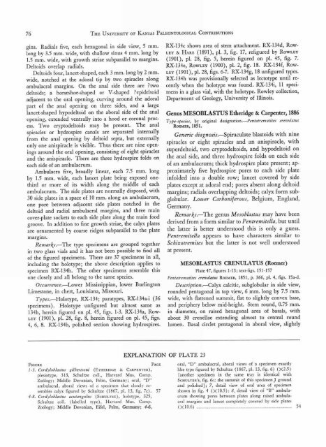
EXPLANATION OF PLATE 23<br />
FIGURE<br />
PAGE<br />
1 -3. Cordyloblastus gilbertsoni (ETHERIDGE & CARPENTER),<br />
plesiotype, 313, Schultze coll., Harvard Mus. Comp.<br />
Zoology; Middle Devonian, Pelm, Germany; oral, "D"<br />
ambulacral, aboral views <strong>of</strong> a specimen that closely resembles<br />
calyx figured by Schultze (1867, pl. 13, fig. 7c). 57<br />
4-8. Cordyloblastus acutangtdus (ScHuurzE), holotype, 325,<br />
Schultze coll. (labelled type), Harvard Mus. Comp.<br />
Zoology; Middle Devonian, Eifel, Pelm, Germany; 4-6,<br />
oral, "D" ambulacral, aboral views <strong>of</strong> a specimen exactly<br />
like type figured by Schultze (1867, pl. 13, fig. 6) ( X2.5)<br />
[another specimen in the same tray is identical with<br />
Scum..-rzE's, fig. 6c; the summit <strong>of</strong> this specimen .1 ground<br />
and polished]; 7, detail view <strong>of</strong> oral area <strong>of</strong> specimen<br />
shown in fig. 4 ( X10.5); 8, detail view <strong>of</strong> "B" ambulacrum<br />
showing pores between plates along raised ambulacral<br />
margins and lancet completely covered by side plates<br />
( X 10.6) 54