ECHINODERMATA - KU ScholarWorks - University of Kansas
ECHINODERMATA - KU ScholarWorks - University of Kansas
ECHINODERMATA - KU ScholarWorks - University of Kansas

You also want an ePaper? Increase the reach of your titles
YUMPU automatically turns print PDFs into web optimized ePapers that Google loves.
56 THE UNIVERSITY OF KANSAS PALEONTOLOGICAL CONTRIBUTIONS<br />
broken. Basal circlet conical in side view, rounded pentagonal<br />
in basal view, 9 mm. long by 7 mm. wide. Radials<br />
five, wide, subquadrangular, each 10 mm. long by 6 mm.<br />
wide, with short, narrow, shallow sinus 6 mm. long by<br />
1.5 mm. wide, with outward-flaring radial lips, giving a<br />
pentalobate appearance to the calyx in top view. The<br />
radial limbs are shorter on the anal side than on the other<br />
sides. Radials overlap deltoids.<br />
Deltoids four, not visible in side view, each divided<br />
into a small quadrangular deltoid body 0.5 mm. long by<br />
0.5 mm. wide and a lip that is approximately 0.5 mm.<br />
wide, with a large, wide oval spiracle between, each<br />
spiracle bordered laterally by a lancet stipe and side<br />
plates. On the anal side there are four deltoid plates,<br />
two <strong>of</strong> which (superdeltoid and hypodeltoid) are seen<br />
externally, the two cryptodeltoids being hidden beneath<br />
the hypodeltoid. The superdeltoid is adjacent to the oral<br />
opening and bordered aborally on each side by a lancet<br />
stipe. The wide oval anispiracle is located between the<br />
superdeltoid and pentagonal hypodeltoid, the latter plate<br />
being 1.5 mm. long by 1.5 mm. wide and barely visible in<br />
side view. Internally, the two cryptodeltoids rest on the<br />
aboral face <strong>of</strong> the superdeltoid, separating the anal spiracles<br />
from the anal opening internally and they are overlapped<br />
by the radial limbs and hypodeltoid. Thus, there<br />
are five openings around the oral opening, the four<br />
spiracles and anispiracle.<br />
Ambulacra five, linear, each 8 mm. long by 1.5 mm.<br />
wide, with lancet covered by side plates except along the<br />
main food groove at the adorai end. There are 22 side<br />
plates in a space <strong>of</strong> 10 mm. along an ambulacrum, one<br />
pore between adjacent side plates along radial margins,<br />
and pores absent along deltoid margins. In mature specimens<br />
there are nine hydrospire folds on each side <strong>of</strong> an<br />
ambulacrum, including the anal side. The surfaces <strong>of</strong><br />
the calyx plates are ornamented with fine growth striae<br />
parallel to plate margins. One abnormal specimen has a<br />
sixth short radial developed on the summit between the<br />
(D) and (E) radials.<br />
There is much variation in this species and more work<br />
needs to be done on the small specimens. In one small<br />
specimen there appeared to be only four hydrospire folds<br />
on each side <strong>of</strong> an ambulacrum.<br />
Occurrence.—Middle Devonian, Eifelian, Pram,<br />
Gerolstein, Nollenbach, Germany.<br />
Types.—Topotypes, 213, one specimen, Schultze collection,<br />
old no. 25, Eifel; 305, six specimens, old no. 2348,<br />
Nollenbach bei Kerpen, Schultze collection. Plesiotype,<br />
195, one specimen, old no. 26, Eifel, Schultze collection,<br />
Harvard Museum <strong>of</strong> Comparative Zoology, Cambridge,<br />
Mass. Plesiotypes, E21,087, two specimens, Eifel and<br />
E21,109, two specimens, Gerolstein, Prussia, Buffalo Society<br />
<strong>of</strong> Natural Sciences, Buffalo, New York. Others not<br />
studied in detail or figured are in the Walker Museum,<br />
87<br />
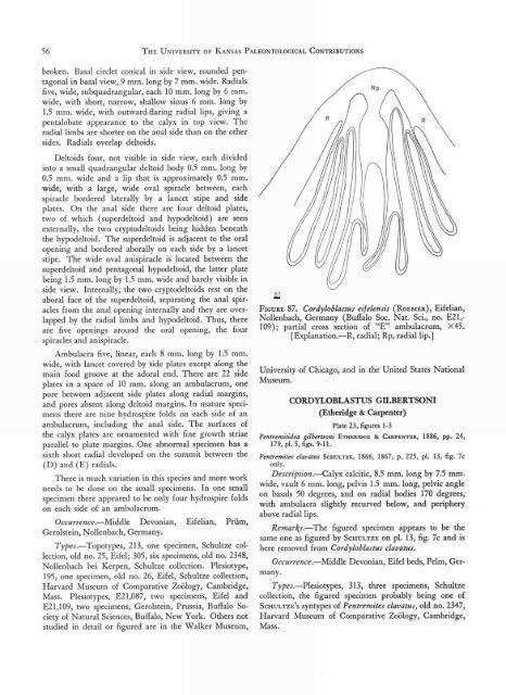
FIGURE 87. Cordyloblastus eifelensis (RoEmEa), Eifelian,<br />
Nollenbach, Germany (Buffalo Soc. Nat. Sci., no. E21,-<br />
109); partial cross section <strong>of</strong> "E" ambulacrum, X45.<br />
[Explanation.—R, radial; Rp, radial lip.]<br />
<strong>University</strong> <strong>of</strong> Chicago, and in the United States National<br />
Museum.<br />
CORDYLOBLASTUS GILBERTSON'<br />
(Etheridge & Carpenter)<br />
Plate 23, figures 1-3<br />
Pentremitidea gilbertsoni ETHERIDGE & CARPENTER, 1886, pp. 24,<br />
179, pl. 5, figs. 9-11.<br />
Pentremites clavatus SCHULTZE, 1866, 1867, p. 225, pl. 13, fig. 7c<br />
only.<br />
Description.—Calyx calcitic, 8.5 mm. long by 7.5 mm.<br />
wide, vault 6 mm. long, pelvis 1.5 mm. long, pelvic angle<br />
on basals 50 degrees, and on radial bodies 170 degrees,<br />
with ambulacra slightly recurved below, and periphery<br />
above radial lips.<br />
Remarks.—The figured specimen appears to be the<br />
same one as figured by SCHULTZE on pl. 13, fig. 7c and is<br />
here removed from Cord yloblastus clavatus.<br />
Occurrence.—Middle Devonian, Eifel beds, Pelm, Germany.<br />
Types.—Plesiotypes, 313, three specimens, Schultze<br />
collection, the figured specimen probably being one <strong>of</strong><br />
SCHULTZE ' S syntypes <strong>of</strong> Pentremites clavatus, old no. 2347,<br />
Harvard Museum <strong>of</strong> Comparative Zoology, Cambridge,<br />
Mass.