ECHINODERMATA - KU ScholarWorks - University of Kansas
ECHINODERMATA - KU ScholarWorks - University of Kansas
ECHINODERMATA - KU ScholarWorks - University of Kansas

Create successful ePaper yourself
Turn your PDF publications into a flip-book with our unique Google optimized e-Paper software.
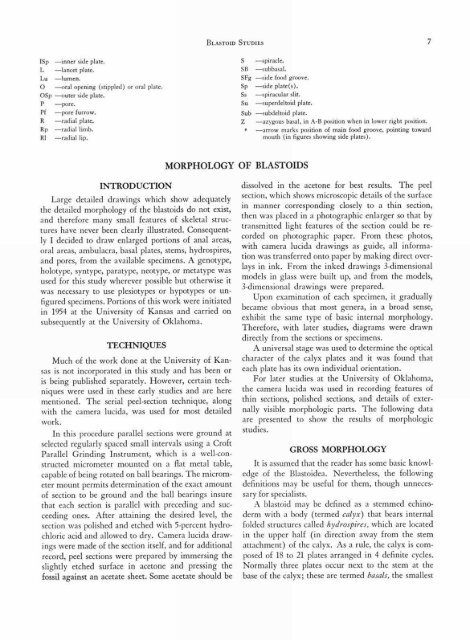
BLASTOID STUDIES 7<br />
ISp —inner side plate.<br />
—lancet plate.<br />
Lu —lumen.<br />
0 —oral opening (stippled) or oral plate.<br />
OSp —outer side plate.<br />
— pore.<br />
Pf —pore furrow.<br />
—radial plate.<br />
Rp —radial limb.<br />
RI —radial lip.<br />
S —spiracle.<br />
SB —subbasal.<br />
SFg —side food groove.<br />
Sp —side plate(s).<br />
Ss —spiracular slit.<br />
Su —superdeltoid plate.<br />
Sub —subdeltoid plate.<br />
Z —azygous basal, in A-B position when in lower right position.<br />
—arrow marks position <strong>of</strong> main food groove, pointing toward<br />
mouth (in figures showing side plates).<br />
MORPHOLOGY OF BLASTOIDS<br />
INTRODUCTION<br />
Large detailed drawings which show adequately<br />
the detailed morphology <strong>of</strong> the blastoids do not exist,<br />
and therefore many small features <strong>of</strong> skeletal structures<br />
have never been clearly illustrated. Consequently<br />
I decided to draw enlarged portions <strong>of</strong> anal areas,<br />
oral areas, ambulacra, basal plates, stems, hydrospires,<br />
and pores, from the available specimens. A genotype,<br />
holotype, syntype, paratype, neotype, or metatype was<br />
used for this study wherever possible but otherwise it<br />
was necessary to use plesiotypes or hypotypes or unfigured<br />
specimens. Portions <strong>of</strong> this work were initiated<br />
in 1954 at the <strong>University</strong> <strong>of</strong> <strong>Kansas</strong> and carried on<br />
subsequently at the <strong>University</strong> <strong>of</strong> Oklahoma.<br />
TECHNIQUES<br />
Much <strong>of</strong> the work done at the <strong>University</strong> <strong>of</strong> <strong>Kansas</strong><br />
is not incorporated in this study and has been or<br />
is being published separately. However, certain techniques<br />
were used in these early studies and are here<br />
mentioned. The serial peel-section technique, along<br />
with the camera lucida, was used for most detailed<br />
work.<br />
In this procedure parallel sections were ground at<br />
selected regularly spaced small intervals using a Cr<strong>of</strong>t<br />
Parallel Grinding Instrument, which is a well-constructed<br />
micrometer mounted on a flat metal table,<br />
capable <strong>of</strong> being rotated on ball bearings. The micrometer<br />
mount permits determination <strong>of</strong> the exact amount<br />
<strong>of</strong> section to be ground and the ball bearings insure<br />
that each section is parallel with preceding and succeeding<br />
ones. After attaining the desired level, the<br />
section was polished and etched with 5-percent hydrochloric<br />
acid and allowed to dry. Camera lucida drawings<br />
were made <strong>of</strong> the section itself, and for additional<br />
record, peel sections were prepared by immersing the<br />
slightly etched surface in acetone and pressing the<br />
fossil against an acetate sheet. Some acetate should be<br />
dissolved in the acetone for best results. The peel<br />
section, which shows microscopic details <strong>of</strong> the surface<br />
in manner corresponding closely to a thin section,<br />
then was placed in a photographic enlarger so that by<br />
transmitted light features <strong>of</strong> the section could be recorded<br />
on photographic paper. From these photos,<br />
with camera lucida drawings as guide, all information<br />
was transferred onto paper by making direct overlays<br />
in ink. From the inked drawings 3-dimensional<br />
models in glass were built up, and from the models,<br />
3-dimensional drawings were prepared.<br />
Upon examination <strong>of</strong> each specimen, it gradually<br />
became obvious that most genera, in a broad sense,<br />
exhibit the same type <strong>of</strong> basic internal morphology.<br />
Therefore, with later studies, diagrams were drawn<br />
directly from the sections or specimens.<br />
A universal stage was used to determine the optical<br />
character <strong>of</strong> the calyx plates and it was found that<br />
each plate has its own individual orientation.<br />
For later studies at the <strong>University</strong> <strong>of</strong> Oklahoma,<br />
the camera lucida was used in recording features <strong>of</strong><br />
thin sections, polished sections, and details <strong>of</strong> externally<br />
visible morphologic parts. The following data<br />
are presented to show the results <strong>of</strong> morphologic<br />
studies.<br />
GROSS MORPHOLOGY<br />
It is assumed that the reader has some basic knowledge<br />
<strong>of</strong> the Blastoidea. Nevertheless, the following<br />
definitions may be useful for them, though unnecessary<br />
for specialists.<br />
A blastoid may be defined as a stemmed echinoderm<br />
with a body (termed calyx) that bears internal<br />
folded structures called hydrospires, which are located<br />
in the upper half (in direction away from the stem<br />
attachment) <strong>of</strong> the calyx. As a rule, the calyx is composed<br />
<strong>of</strong> 18 to 21 plates arranged in 4 definite cycles.<br />
Normally three plates occur next to the stem at the<br />
base <strong>of</strong> the calyx; these are termed basais, the smallest