62 THE UNIVERSITY OF KANSAS PALEONTOLOGICAL CONTRIBUTIONS DEVONOBLASTUS LEDA (Hall) Plate 24, figures 1-2, 6 Pentrenntes leda HALL, 1862, p. 149, pl. 1, fig. 11. Devonoblastus leda REIMANN, 1935, p. 31. Description.—A detailed description <strong>of</strong> the syntypes has been submitted for publication in another work. The syntypes are not well preserved and most <strong>of</strong> the generic information is taken from the description <strong>of</strong> specimens <strong>of</strong> Devonoblastus whiteavesi. The syntypes are figured on Plate 24. This species differs from D. whiteavesi in that the maximum width (or periphery) <strong>of</strong> D. leda is low, at or near the radial lips, in contrast to its higher, more median position in D. whiteavesi. Occurrence.—Devonian, Hamilton Shale, western New York. Types.—Syntypes, 451 (figures 1, 6), and 452 (figure 2), two specimens, Hall collection, New York State Museum, Albany, New York. DEVONOBLASTUS WHITEAVESI Reimann Plate 24, figures 3-5; plate 25, figures 1-12; text-figs. 101-112 Devonoblastus whiteavesi REIMANN, 1935, p. 32, pl. 1, fig. 8. Granatocrinus leda WHITEAVES, 1889, p. 108, pl. 14, fig. 14. Description.—The description is mainly on specimen 35,036, supplemented by information from other specimens. The calyx is calcitic, oval in side view, with short conical pelvis, 16 mm. long by 10.5 mm. wide, pentagonal in top view, with periphery near mid-height, and vault 14 mm. long, pelvis 2 mm. long, pelvic angle on basais 85 degrees and on radial bodies 130 degrees. The stem is round, crenellar, 1.5 mm. in diameter, with approximately 45 crenellae extending inward radially from the margin one-third <strong>of</strong> the distance to the small round lumen. The stem is attached to the basal circlet, which has a hexagonal raised area at the aboral ends <strong>of</strong> the basal plates. Basal circlet pentagonal in aboral view, widely conical in side view, 2 mm. long by 5 mm. wide, with strongly rounded medial ridge on each basal. Radials five, elongate hexagonal, each 14.5 mm. long by 5.5 mm. wide, with long, narrow, shallow sinus 13 mm. long by 2 mm. wide; radials overlapping deltoids. The surfaces <strong>of</strong> the radials and basais are ornamented by fine growth striae parallel to plate margins. Deltoids four, lancet-shaped, each 3.5 mm. long by 2 mm. wide, visible in side view, with one large oval spiracle notched in the adorai end <strong>of</strong> each, separating the deltoid into two parts, the deltoid lip located adjacent to the oral opening and the deltoid body adjacent to the radial limbs. The surfaces <strong>of</strong> the deltoid bodies are ornamented by coarse growth ridges parallel to plate margins, especially parallel to the radiodeltoid sutures. On the anal side four anal deltoid plates seem to be present. The small pentagonal superdeltoid is adjacent to the oral opening and forms the external border on the adorai part <strong>of</strong> the large oval anispiracle. Internally the two cryptodeltoids rest on the aboral face <strong>of</strong> the superdeltoid and separate the anal opening from the adjacent hydrospire canals. The cryptodeltoids are infolded to form hydrospires on the anal side, and are overlapped by the radial limbs. The cryptodeltoids are covered externally by the hypodeltoid plate and are not visible from the exterior. The pentagonal hypodeltoid is visible in side view and is homologous with the deltoid body <strong>of</strong> each <strong>of</strong> the other four deltoids. Five hydrospire folds are seen on each side <strong>of</strong> an ambulacrum. Ambulacra five, linear, slightly recurved below periphery, each 16 mm. long by 2 mm. wide, with lancet covered by side plates except at the adorai end, 24 side plates in a space <strong>of</strong> 10 mm. along an ambulacrum. The side plates are normally disposed, with four main cover-plate lobes to each side plate along the main food groove. A single large pore is observed between adjacent side plates along the radial ambulacral margins, and one or two pores occur along the deltoid ambulacral margins just above the radiodeltoid sutures. Each spiracle is bordered by a deltoid lip, two lancet stipes, two side plates, and a deltoid body. Occurrence.—Middle Devonian, coral zone <strong>of</strong> Widder beds, Hungry Hollow Formation, Thedford and Arkona areas, Ontario. Shale layer <strong>of</strong> Tichenor Limestone, Cazenovia Creek, Springbrook, Erie County, New York. Types.—Topotype, 35,036, one specimen, Charles Southworth collection, from Ausable River valley, 2.5 miles northeast <strong>of</strong> Arkona; <strong>University</strong> <strong>of</strong> Michigan, Ann Arbor. Plesiotype, 34,467, one specimen, from tile yard 0.5 mile north <strong>of</strong> Thedford, Ontario, Charles Southworth collection (June, 1954), <strong>University</strong> <strong>of</strong> Michigan. Plesiotypes, E13,065, three specimens, Charles Southworth collection (1941), from area near Thedford, Ontario, Buffalo Society <strong>of</strong> Natural Sciences, Buffalo, New York. Hypotype, 3,661, one specimen, from Thedford area, described by WHITEAVES (1889) as Granatocrinus leda, Geological Survey <strong>of</strong> Canada, Ottawa, Ontario, Canada. Plesiotypes, S4,648, two specimens, Springer collection, from Ausable River valley, 2 miles east <strong>of</strong> Arkona, Ontario, collected by Charles Southworth; S4,144, one specimen, Springer collection, from Marsh's Mill, Arkona, Ontario, lower Widder beds; and 142,025, one specimen, labelled Devonoblastus leda, Tichenor Limestone, New York, all in U. S. National Museum, Washington. Genus DEPLOBLASTUS Fay, n. gen. Type-spedes, by original designation (herein).—Granatocrinus glaber MEEK & WORTHEN, 1869. Generic diagnosis.—Spiraculate blastoids with five paired spiracles, or four paired spiracles in addition to a paired anispiracle, with superdeltoid, two cryptodeltoids, and hypodeltoid on anal side; two hydrospire folds on each side <strong>of</strong> an ambulacrum; lancet covered
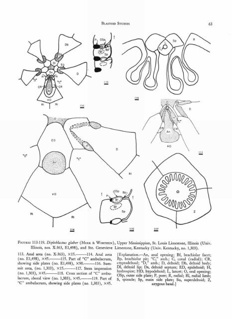
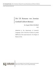
BLASTOID STUDIES 63 FIGURES 113-119. Diploblastus glaber (MEEK & WORTHEN), Upper Mississippian, St. Louis Limestone, Illinois (Univ. Illinois, nos. X-363, E1,498), and Ste. Genevieve Limestone, Kentucky (Univ. Kentucky, no. 1,303). 113. Anal area (no. X-363), X15. 114. Anal area (no. E1,498), X45. 115. Part <strong>of</strong> "C" ambulacrum, showing side plates (no. E1,498), X90. 116. Summit area, (no. 1,303), X15. 117. Stem impression (no. 1,303), X45. 118. Cross section <strong>of</strong> "C" ambulacrum, aboral view (no. 1,303), X45.— 119. Part <strong>of</strong> ambulacrum, showing side plates (no 1,303), X45. [Explanation.—An, anal opening; Bf, brachiolar facet; Bp, brachiolar pit; "C," amb.; C, canal (radial); CR, cryptodeltoid; "D," amb.; D, deltoid; Db, deltoid body; D1, deltoid lip; Ds, deltoid septum; ED, epideltoid; H, hydrospire; HD, hypodeltoid; L, lancet; 0, oral opening; OSp, outer side plate; P, pore; R, radial; RI, radial limb; S, spiracle; Sp, main side plate; Su, superdeltoid; Z, azygous basal.]