ECHINODERMATA - KU ScholarWorks - University of Kansas
ECHINODERMATA - KU ScholarWorks - University of Kansas
ECHINODERMATA - KU ScholarWorks - University of Kansas

You also want an ePaper? Increase the reach of your titles
YUMPU automatically turns print PDFs into web optimized ePapers that Google loves.
BLASTOID STUDIES 91<br />
189 191 192<br />
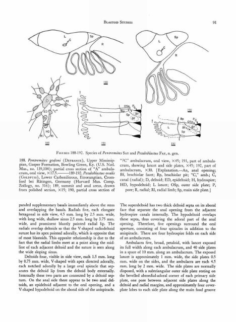
FIGURES 188-192. Species <strong>of</strong> Pentremites SAY and Petaloblastus FAY, n. gen.<br />
188. Pentremites godoni (DEFRANcE), Upper Mississippian,<br />
Gasper Formation, Bowling Green, Ky. (U.S. Natl.<br />
Mus., no. 139,104); partial cross section <strong>of</strong> "A" ambulacrum,<br />
oral view, X17.5. 189-192. Petaloblastus ovalis<br />
(GoLD Fuss), Lower Carboniferous, Etroeungtian, Cromford<br />
bei Ratingen, Germany (Harvard Mus. Comp.<br />
Zoology, no. 316); 189, summit and anal areas, drawn<br />
from polished section, X15; 190, partial cross section <strong>of</strong><br />
"?C" ambulacrum, oral view, X45; 191, part <strong>of</strong> ambulacrum,<br />
showing lancet and side plates, X45; 192, part <strong>of</strong><br />
ambulacrum, X30. [Explanation.—An, anal opening;<br />
Bf, brachiolar facet; Bp, brachiolar pit; "C," amb.; C,<br />
canal (radial); D, deltoid; ED, epideltoid; H, hydrospire;<br />
HD, hypodeltoid; L lancet; OSp, outer side plate; P,<br />
pore; R, radial; RI, radial limb; Sp, main side plate.]<br />
panded supplementary basais immediately above the stem<br />
and overlapping the basals. Radials five, each elongate<br />
hexagonal in side view, 4.5 mm. long by 2.5 mm. wide,<br />
with long wide, shallow sinus 2.5 mm. long by 3.75 mm.<br />
wide, and prominent bluntly pointed radial lip. The<br />
radials overlap deltoids so that the V-shaped radiodeltoid<br />
suture has its apex pointed adorally, which is opposite that<br />
<strong>of</strong> most blastoids. This opposite relationship is due to the<br />
fact that the radial limbs meet at a point along the midline<br />
<strong>of</strong> each adjacent deltoid and the suture is seen along<br />
the wide sloping sinus.<br />
Deltoids four, visible in side view, each 1.5 mm. long<br />
by 0.75 mm. wide, V-shaped with apex directed adorally,<br />
each notched adorally by a large oval spiracle that separates<br />
the deltoid lip from the deltoid body externally.<br />
Internally these two parts are connected by a deltoid septum.<br />
On the anal side there appear to be two anal deltoids,<br />
an epideltoid adjacent to the oral opening, and a<br />
V-shaped hypodeltoid on the aboral side <strong>of</strong> the anispiracle.<br />
The superdeltoid has two thick deltoid septa on its aboral<br />
face that separate the anal opening from the adjacent<br />
hydrospire canals internally. The hypodeltoid overlaps<br />
these septa, thus covering the adoral part <strong>of</strong> the anal<br />
opening. Therefore, five openings surround the oral<br />
aperture, consisting <strong>of</strong> four spiracles in addition to the<br />
anispiracle. There are four hydrospire folds on each side<br />
<strong>of</strong> an ambulacrum.<br />
Ambulacra five, broad, petaloid, with lancet exposed<br />
its full width along each ambulacrum, and 40 side plates<br />
in a space <strong>of</strong> 10 mm. along an ambulacrum. The exposed<br />
lancet is approximately 1 mm. wide, the side plates 0.5<br />
mm. wide on the sides, and the ambulacra are each 4.5<br />
mm. long by 2 mm. wide. The side plates are normally<br />
disposed, with a subtriangular outer side plate resting on<br />
the bevelled abmedial-adoral corner <strong>of</strong> each primary side<br />
plate, one pore between adjacent side plates along the<br />
deltoid and radial margins, and approximately four coverplate<br />
lobes to each side plate along the main food groove