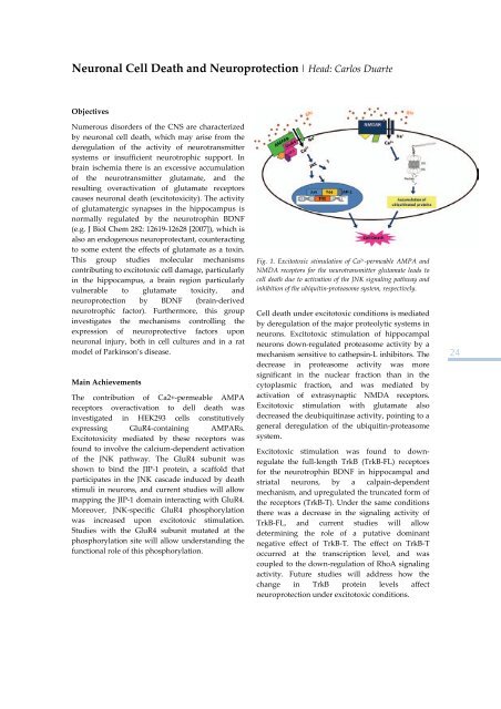
Neuronal Cell Death <strong>and</strong> Neuroprotection | Head: Carlos DuarteObjectivesNumerous disorders <strong>of</strong> the CNS are characterizedby neuronal cell death, which may arise from thederegulation <strong>of</strong> the activity <strong>of</strong> neurotransmittersystems or insufficient neurotrophic support. Inbrain ischemia there is an excessive accumulation<strong>of</strong> the neurotransmitter glutamate, <strong>and</strong> theresulting overactivation <strong>of</strong> glutamate receptorscauses neuronal death (excitotoxicity). The activity<strong>of</strong> glutamatergic synapses in the hippocampus isnormally regulated by the neurotrophin BDNF(e.g. J Biol Chem 282: 12619‐12628 [2007]), which isalso an endogenous neuroprotectant, counteractingto some extent the effects <strong>of</strong> glutamate as a toxin.This group studies molecular mechanismscontributing to excitotoxic cell damage, particularlyin the hippocampus, a brain region particularlyvulnerable to glutamate toxicity, <strong>and</strong>neuroprotection by BDNF (brain‐derivedneurotrophic factor). Furthermore, this groupinvestigates the mechanisms controlling theexpression <strong>of</strong> neuroprotective factors uponneuronal injury, both in cell cultures <strong>and</strong> in a ratmodel <strong>of</strong> Parkinson’s disease.Main AchievementsThe contribution <strong>of</strong> Ca2+‐permeable AMPAreceptors overactivation to dell death wasinvestigated in HEK293 cells constitutivelyexpressing GluR4‐containing AMPARs.Excitotoxicity mediated by these receptors wasfound to involve the calcium‐dependent activation<strong>of</strong> the JNK pathway. The GluR4 subunit wasshown to bind the JIP‐1 protein, a scaffold thatparticipates in the JNK cascade induced by deathstimuli in neurons, <strong>and</strong> current studies will allowmapping the JIP‐1 domain interacting with GluR4.Moreover, JNK‐specific GluR4 phosphorylationwas increased upon excitotoxic stimulation.Studies with the GluR4 subunit mutated at thephosphorylation site will allow underst<strong>and</strong>ing thefunctional role <strong>of</strong> this phosphorylation.Fig. 1. Excitotoxic stimulation <strong>of</strong> Ca 2+ ‐permeable AMPA <strong>and</strong>NMDA receptors <strong>for</strong> the neurotransmitter glutamate leads tocell death due to activation <strong>of</strong> the JNK signaling pathway <strong>and</strong>inhibition <strong>of</strong> the ubiquitin‐proteasome system, respectively.Cell death under excitotoxic conditions is mediatedby deregulation <strong>of</strong> the major proteolytic systems inneurons. Excitotoxic stimulation <strong>of</strong> hippocampalneurons down‐regulated proteasome activity by amechanism sensitive to cathepsin‐L inhibitors. Thedecrease in proteasome activity was moresignificant in the nuclear fraction than in thecytoplasmic fraction, <strong>and</strong> was mediated byactivation <strong>of</strong> extrasynaptic NMDA receptors.Excitotoxic stimulation with glutamate alsodecreased the deubiquitinase activity, pointing to ageneral deregulation <strong>of</strong> the ubiquitin‐proteasomesystem.Excitotoxic stimulation was found to downregulatethe full‐length TrkB (TrkB‐FL) receptors<strong>for</strong> the neurotrophin BDNF in hippocampal <strong>and</strong>striatal neurons, by a calpain‐dependentmechanism, <strong>and</strong> upregulated the truncated <strong>for</strong>m <strong>of</strong>the receptors (TrkB‐T). Under the same conditionsthere was a decrease in the signaling activity <strong>of</strong>TrkB‐FL, <strong>and</strong> current studies will allowdetermining the role <strong>of</strong> a putative dominantnegative effect <strong>of</strong> TrkB‐T. The effect on TrkB‐Toccurred at the transcription level, <strong>and</strong> wascoupled to the down‐regulation <strong>of</strong> RhoA signalingactivity. Future studies will address how thechange in TrkB protein levels affectneuroprotection under excitotoxic conditions.24
Mitochondrial Dysfuntion <strong>and</strong> Cell Death | Head: Ana Cristina RegoObjectivesSelective neurodegeneration in irreversible disorders<strong>of</strong> the CNS has been largely attributed to proteinmisfolding, excitotoxicity <strong>and</strong> mitochondrialimpairment. However, how modified or mutantproteins interfere with mitochondrial <strong>and</strong> neuronalfunction is largely unclear. Furthermore, severalneurotoxic substances cause neuronal death throughchanges in mitochondrial activity, deregulation <strong>of</strong>intracellular calcium homeostasis <strong>and</strong> oxidativestress. Thus, the main objective <strong>of</strong> our group is tostudy defective intracellular signaling mechanisms<strong>and</strong> identify molecular targets <strong>for</strong> therapeuticintervention underlying excitotoxicity, mitochondrialdysfunction, oxidative stress <strong>and</strong> neuronal death inneurodegenerative diseases, including polyglutamineexpansion (Huntington’s (HD) <strong>and</strong> Machado‐Joseph’s (MJD) disorders), Parkinson’s (PD) <strong>and</strong>Alzheimer’s (AD) diseases, <strong>and</strong> in the neuropathologycaused by drug addiction. We also aim to evaluatethe efficacy <strong>of</strong> neuroprotective strategies (pharmacological<strong>and</strong> cell replacement therapies) that help recoveringcell function <strong>and</strong> thus cell survival in animal <strong>and</strong>cellular models <strong>of</strong> neurodegenerative disorders.The protective effect <strong>of</strong> growth factors that act astrophic signalling molecules assumes a highimportance in neurodegeneration. There<strong>for</strong>e, wewill examine the influence <strong>of</strong> brain‐derivedneurotrophic (BDNF) <strong>and</strong> glial cell‐derivedneurotrophic factor (GDNF) either directly or aftertransduction into stem cells acquiring a neuralphenotype, <strong>and</strong> the neuroprotective effect <strong>of</strong>insulin‐growth factor (IGF‐1), insulin <strong>and</strong> histonedeacetylase modulators in PD <strong>and</strong> HD models.derived from HD knock‐in mice. Mutant cellsrevealed increased ROS <strong>for</strong>mation, decreased GSHsynthesis <strong>and</strong> in the activity <strong>of</strong> antioxidantenzymes. We further studied markers <strong>of</strong> apoptosis<strong>and</strong> mitochondrial dysfunction in peripheral bloodcells <strong>of</strong> HD patients (Almeida et al, <strong>2008</strong>). Wefound increased Bax expression in B <strong>and</strong> Tlymphocytes, <strong>and</strong> monocytes from HD patients. Blymphocytes also showed decreased mitochondrialpotential, suggesting that these cells may reflectchanges observed in HD brain. In the context <strong>of</strong>MJD, we observed an increase in ataxin‐3deubiquitinating activity in vitro after long periods<strong>of</strong> incubation with valosin‐containing protein(VCP/p97). Using human SH‐SY5Y cells transfectedwith WT <strong>and</strong> mutant A53T alpha‐synuclein (a‐syn)subjected to iron <strong>and</strong> rotenone as models <strong>of</strong> PD, wedetected increased P‐Ser129‐a‐syn, decreasedmitochondrial potential <strong>and</strong> ROS production, inthe absence <strong>of</strong> a‐syn aggregates. Within the scope<strong>of</strong> AD pathogenesis, we observed that amyloidbetapeptide (Abeta) oligomers increase NR2A denovo protein synthesis, but enhance membranesurface <strong>of</strong> NR2B NMDAR subunits in maturehippocampal neurons. Abeta evoked calcium risesensitive to NMDAR antagonists <strong>and</strong> decreasedtubulin‐beta III levels, suggesting the involvement<strong>of</strong> NMDAR subunits on neuronal dysfunctioncaused by Abeta oligomers. Regarding research ondrugs <strong>of</strong> abuse we showed that combination <strong>of</strong>cocaine <strong>and</strong> heroin enhances the neurotoxicity <strong>of</strong>the drugs alone <strong>and</strong> that cocaine‐morphine adductsshift cell death pathways towards necrosis.25Main AchievementsIn the context <strong>of</strong> HD, our group showed thatactivation <strong>of</strong> BDNF signaling pathways prevent 3‐NP toxicity <strong>and</strong> transcriptional deregulation inneurons. We also observed alterations in themitochondrial proteome <strong>of</strong> HD mice brain <strong>and</strong> incomplex I activity, which were prevented byBDNF. These data implicate expression <strong>of</strong>mitochondrial proteins <strong>and</strong> molecular activity inthis organelle as HD therapeutic targets. Moreover,we demonstrated insulin neuroprotection againstoxidative stress through increased expression <strong>of</strong>proteins involved in antioxidant defense, glucosemetabolism <strong>and</strong> prevention against apoptosis(Duarte et al, <strong>2008</strong>). We also observed insulinprotection against oxidative stress in striatal cellsInsulin‐mediated mRNA expression <strong>of</strong> glutathione peroxidase‐1 (A), hexokinase‐II (B), Bcl‐2 (C) <strong>and</strong> caspase‐3 (D) uponoxidative stress in cultured cortical neurons labeling theinsulin receptor (IR) (E) [Duarte et al., <strong>2008</strong>].
- Page 7 and 8: General ObjectivesThe CNC major mis
- Page 11 and 12: OrganizationThe Center for Neurosci
- Page 13: Microbiology | Milton CostaMicrobio
- Page 16 and 17: per year) will be proposed by the g
- Page 18 and 19: Neuroprotection and Neurogenesis in
- Page 20 and 21: Retinal Dysfunction and Neurogenesi
- Page 22 and 23: Glutamatergic synapses | Head: Ana
- Page 26 and 27: Molecular Mechanisms of Disease | H
- Page 28 and 29: PublicationsAgasse F, Bernardino L,
- Page 30 and 31: Santiago AR, Carvalho, CM, Carvalho
- Page 32 and 33: “Nano‐transportadores de base l
- Page 34 and 35: Vectors and Gene Therapy GroupM. Co
- Page 36 and 37: Molecular Systems Biology | Head: A
- Page 38 and 39: Vectors and Gene Therapy | Head: Ma
- Page 40 and 41: PublicationsAlves S, Nascimento‐F
- Page 43 and 44: Area C | Cell and Molecular Toxicol
- Page 45 and 46: Mitochondrial Toxicology and Pharma
- Page 47: Pharmacometrics GroupAmílcar Falc
- Page 50 and 51: Free Radicals and Antioxidants in B
- Page 52 and 53: Pharmacometrics | Head: Amílcar Fa
- Page 54 and 55: Correia S, Carvalho C, Santos MS, P
- Page 57 and 58: Area D | MicrobiologyCoordinator |
- Page 59: Microbiology of Extreme Environment
- Page 62 and 63: Medical Mycology - Yeast Research |
- Page 65 and 66: Area E | Biophysics and Biomedical
- Page 67 and 68: Inorganic Biochemistry and Molecula
- Page 69 and 70: Inorganic Biochemistry and Molecula
- Page 71 and 72: Cell Biophysics |Head: Luís Martin
- Page 73: Sobral AJFN, Justino LLG, Santos AC
- Page 76 and 77:
Future PlansThere is an enormous we
- Page 78 and 79:
Paula MotaSara M. Diniz Martins Lop
- Page 80 and 81:
Biology of Reproduction and Human F
- Page 82 and 83:
Insulin Resistance and Adipocyte |
- Page 85 and 86:
Biomedical Inter‐Institutional Re
- Page 87 and 88:
3. Pediatric Research: metabolic di
- Page 89 and 90:
PublicationsSantos MJ, Cleto S, Men
- Page 91:
7. Research in brain cancer: geneti
- Page 94 and 95:
Grafting SVZ neural stem cell cultu
- Page 96 and 97:
Structure‐function analysis of th
- Page 98 and 99:
Anticancer Effects of of Phytochemi
- Page 100 and 101:
Investigaciones Biomédicas “Albe
- Page 102 and 103:
Participation in the organization o
- Page 104 and 105:
May 2008Member of the organizing co
- Page 106 and 107:
106
- Page 108 and 109:
Genome BiologyFebruary 25 ‐ 27Isa
- Page 110 and 111:
Seminars2008 Series | CNC Audithori
- Page 112 and 113:
13.6.2008Cells caught in the act: M
- Page 114 and 115:
5.12.2008Unexpected fate and functi
- Page 116 and 117:
Elisabete Ferreiro“Cross‐talk b
- Page 118 and 119:
Ana Isabel Vicente Rafael“Estudos
- Page 120 and 121:
Rosete Pais“Papilomavirus humano
- Page 122 and 123:
122
- Page 124 and 125:
124
- Page 126 and 127:
FLOW CYTOMETRY UNITHead of Unit: Is
- Page 128 and 129:
MASS SPECTROMETRY UNITHead of Unit:
- Page 130 and 131:
130
- Page 132 and 133:
Amino Acid AnalysisOur laboratory r
- Page 134 and 135:
2. Areas of Expertise / Research /
- Page 136 and 137:
Multiple SclerosisAn extension of t
- Page 138 and 139:
2.2. Centre for Bioavailability Stu
- Page 140 and 141:
StaffCoordinatorTice Macedo, MD, Ph
- Page 142 and 143:
142
- Page 144 and 145:
Compatible solutes from extremophil
- Page 146 and 147:
Proteases aspárticas secretadas em
- Page 148 and 149:
Mecanismos de plasticidade sinápti
- Page 150 and 151:
Clivagem dos transportadores vesicu
- Page 152 and 153:
ʺBIOINK ‐ Aprendizagem increment
- Page 154 and 155:
154
- Page 156 and 157:
Henrique Faneca (Auxiliar Inv., CNC
- Page 158 and 159:
Liliana Bernardino 100Luis Miguel E
- Page 160 and 161:
João Teixeira 100João Teodoro 100
- Page 162 and 163:
Patricia Henriques Domingues 100Pat
- Page 164 and 165:
ADMINISTRATIVE STAFFTime % at CNCAr
- Page 166 and 167:
Sandra Isabel M. Cardoso (Auxiliar
- Page 168 and 169:
Molecular Biotechnology and HealthE
- Page 170 and 171:
Cell and Molecular ToxicologyLeonor
- Page 172 and 173:
MicrobiologyMilton Costa, PhD, Coor
- Page 174 and 175:
Paulo Gameiro Guerreiro 100Pedro Co
- Page 176 and 177:
Marta Isabel Rodrigues Baptista 100
- Page 178:
Url: http://www.cnbc.pt | Email: in