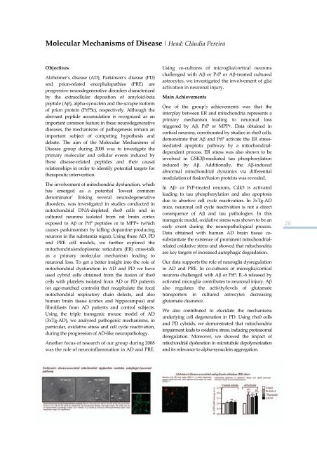
Molecular Mechanisms <strong>of</strong> Disease | Head: Cláudia PereiraObjectivesAlzheimer’s disease (AD), Parkinson’s disease (PD)<strong>and</strong> prion‐related encephalopathies (PRE) areprogressive neurodegenerative disorders characterizedby the extracellular deposition <strong>of</strong> amyloid‐betapeptide (Aβ), alpha‐synuclein <strong>and</strong> the scrapie is<strong>of</strong>orm<strong>of</strong> prion protein (PrPSc), respectively. Although theaberrant peptide accumulation is recognized as animportant common feature in these neurodegenerativediseases, the mechanisms <strong>of</strong> pathogenesis remain animportant subject <strong>of</strong> competing hypothesis <strong>and</strong>debate. The aim <strong>of</strong> the Molecular Mechanisms <strong>of</strong>Disease group during <strong>2008</strong> was to investigate theprimary molecular <strong>and</strong> cellular events induced bythese disease‐related peptides <strong>and</strong> their causalrelationships in order to identify potential targets <strong>for</strong>therapeutic intervention.The involvement <strong>of</strong> mitochondria dysfunction, whichhas emerged as a potential ‘lowest commondenominator’ linking several neurodegenerativedisorders, was investigated in studies conducted inmitochondrial DNA‐depleted rho0 cells <strong>and</strong> incultured neurons isolated from rat brain cortexexposed to Aβ or PrP peptides or to MPP+ (whichcauses parkinsonism by killing dopamine‐producingneurons in the substantia nigra). Using these AD, PD<strong>and</strong> PRE cell models, we further explored themitochondria/endoplasmic reticulum (ER) cross‐talkas a primary molecular mechanism leading toneuronal loss. To get a better insight into the role <strong>of</strong>mitochondrial dysfunction in AD <strong>and</strong> PD we haveused cybrid cells obtained from the fusion <strong>of</strong> rho0cells with platelets isolated from AD or PD patients(or age‐matched controls) that recapitulate the focalmitochondrial respiratory chain defects, <strong>and</strong> alsohuman brain tissue (cortex <strong>and</strong> hippocampus) <strong>and</strong>fibroblasts from AD patients <strong>and</strong> control subjects.Using the triple transgenic mouse model <strong>of</strong> AD(3xTg‐AD), we analysed pathogenic mechanisms, inparticular, oxidative stress <strong>and</strong> cell cycle reactivation,during the progression <strong>of</strong> AD‐like neuropathology.Another focus <strong>of</strong> research <strong>of</strong> our group during <strong>2008</strong>was the role <strong>of</strong> neuroinflammation in AD <strong>and</strong> PRE.Using co‐cultures <strong>of</strong> microglia/cortical neuronschallenged with Aβ or PrP or Aβ‐treated culturedastrocytes, we investigated the involvement <strong>of</strong> gliaactivation in neuronal injury.Main AchievementsOne <strong>of</strong> the group’s achievements was that theinterplay between ER <strong>and</strong> mitochondria represents aprimary mechanism leading to neuronal losstriggered by Aβ, PrP or MPP+. Data obtained incortical neurons, corroborated by studies in rho0 cells,demonstrate that Aβ <strong>and</strong> PrP activate the ER stressmediatedapoptotic pathway by a mitochondrialdependentprocess. ER stress was also shown to beinvolved in GSK3β‐mediated tau phosphorylationinduced by Aβ. Additionally, the Aβ‐inducedabnormal mitochondrial dynamics via differentialmodulation <strong>of</strong> fission/fusion proteins was revealed.In Aβ‐ or PrP‐treated neurons, Cdk5 is activatedleading to tau phosphorylation <strong>and</strong> also apoptosisdue to abortive cell cycle reactivation. In 3xTg‐ADmice, neuronal cell cycle reactivation is not a directconsequence <strong>of</strong> Aβ <strong>and</strong> tau pathologies. In thistransgenic model, oxidative stress was shown to be anearly event during the neuropathological process.Data obtained with human AD brain tissue cosubstantiatethe existence <strong>of</strong> prominent mitochondrialrelatedoxidative stress <strong>and</strong> showed that mitochondriaare key targets <strong>of</strong> increased autophagic degradation.Our data supports the role <strong>of</strong> neuroglia dysregulationin AD <strong>and</strong> PRE. In co‐cultures <strong>of</strong> microglia/corticalneurons challenged with Aβ or PrP, IL‐6 released byactivated microglia contributes to neuronal injury. Aβalso regulates the activity/levels <strong>of</strong> glutamatetransporters in cultured astrocytes decreasingglutamate clearance.We also contributed to elucidate the mechanismsunderlying cell degeneration in PD. Using rho0 cells<strong>and</strong> PD cybrids, we demonstrated that mitochondriaimpairment leads to oxidative stress, inducing proteasomalderegulation. Moreover, we showed the impact <strong>of</strong>mitochondrial dysfunction in microtubule depolymerization<strong>and</strong> its relevance to alpha‐synuclein aggregation.26
Retinal Dysfunction <strong>and</strong> Neurogenesis | Head: Claudia CavadasObjectivesThe retina is a neuronal structure highlysusceptible to several insults, such ashyperglycemia, excitotoxicity, inflammation <strong>and</strong>exposure to drugs. Our group is activelycommitted to identify important players <strong>and</strong>mechanisms mediating retinal <strong>and</strong> neuronaldamage, with the main goal <strong>of</strong> devising newtherapeutic targets <strong>and</strong> strategies to treat retinal orbrain degenerative diseasesWe will clarify the role(s) <strong>of</strong> NPY in retinalphysiology, <strong>and</strong> further investigate the potentialneuroprotective <strong>and</strong> regulatory effects <strong>of</strong> NPY onretinal progenitor cell proliferation <strong>and</strong>differentiation.We will continue to study the impact <strong>of</strong>hyperglycemia in the retina, namely on exocytosisevents, on the molecular mechanisms underlyingchanges in AMPA receptor subunits expression inretinal cells, <strong>and</strong> on the regulation <strong>of</strong> retinalmicroglia.The evaluation <strong>of</strong> the potential harmful effect <strong>of</strong>ecstasy in rat retinal physiology <strong>and</strong> morphology isanother goal <strong>of</strong> our group.The identification <strong>of</strong> the signaling pathways <strong>and</strong>molecular mechanisms responsible <strong>for</strong> theproliferative/antiproliferative effects <strong>of</strong> nitric oxideon neural stem cells is another main researchinterest <strong>of</strong> our group.In a project <strong>of</strong> close cooperation with thepharmaceutical industry, we are evaluating theneurotoxicity/safety pr<strong>of</strong>ile <strong>of</strong> eslicarbazepineacetate (developed by BIAL, Portugal) <strong>and</strong> itsmetabolites, as compared to other antiepilepticdrugs (AEDs). We are also investigating the effects<strong>of</strong> AEDs on the proliferation <strong>and</strong> fate <strong>of</strong> neuralstem cells.Main AchievementsStarting by the hypothesis that glutamate, the mainexcitatory neurotransmitter in the retina, might beinvolved in the pathogenesis <strong>of</strong> diabeticretinopathy, we found that diabetes or elevatedglucose levels can impair the uptake <strong>and</strong> therelease <strong>of</strong> excitatory neurotransmitters in the retina<strong>and</strong> alter the expression <strong>of</strong> ionotropic glutamatereceptor subunits. In fact, we observed thatdiabetes changes the expression <strong>of</strong> ionotropicglutamate receptor subunits in the human retina,suggesting that glutamatergic transmission in theretina might be compromised early in the course <strong>of</strong>diabetes. Moreover, inflammation, <strong>and</strong> activation<strong>of</strong> microglial cells, has been shown to have animportant role in diabetic retinopathy. We haveshown that high glucose alters the purinergicsignaling system in the retina, resulting high levels<strong>of</strong> extracellular ATP that may lead to inflammationinvolved in the pathogenesis <strong>of</strong> diabeticretinopathy. Moreover, we observed that Mullercells do not influence the adhesion <strong>of</strong> leukocytes toretinal endothelial cellsWe have also described that ecstasy (MDMA)induces retinal cell death, <strong>and</strong> we identifiedneuropeptide Y (NPY) as neuroprotective agentagainst this insult. Moreover, NPY stimulates theproliferation <strong>of</strong> retinal progenitor cells mediated bythe activation <strong>of</strong> NPY Y1, Y2, <strong>and</strong> Y5 receptors aswell as the by the nitric oxide (NO)‐guanylylcyclase pathway. This pathway is also activated byNPY when it stimulates catecholamine release frommouse neuronal‐like cells (chromaffin cells).The study on proliferation <strong>of</strong> endogenous neuralprogenitor cells, as a strategy to promote neuronalrepair, showed that NO stimulates the proliferation<strong>of</strong> neural stem cells by passing the epidermalgrowth factor receptor.Moreover, in a parallel research line, our resultsshow that eslicarbazepine acetate (BIA 2‐093) <strong>and</strong>its metabolites are not toxic to hippocampalneurons compared to carbamazepine oroxcarbazepine.27
- Page 7 and 8: General ObjectivesThe CNC major mis
- Page 11 and 12: OrganizationThe Center for Neurosci
- Page 13: Microbiology | Milton CostaMicrobio
- Page 16 and 17: per year) will be proposed by the g
- Page 18 and 19: Neuroprotection and Neurogenesis in
- Page 20 and 21: Retinal Dysfunction and Neurogenesi
- Page 22 and 23: Glutamatergic synapses | Head: Ana
- Page 24 and 25: Neuronal Cell Death and Neuroprotec
- Page 28 and 29: PublicationsAgasse F, Bernardino L,
- Page 30 and 31: Santiago AR, Carvalho, CM, Carvalho
- Page 32 and 33: “Nano‐transportadores de base l
- Page 34 and 35: Vectors and Gene Therapy GroupM. Co
- Page 36 and 37: Molecular Systems Biology | Head: A
- Page 38 and 39: Vectors and Gene Therapy | Head: Ma
- Page 40 and 41: PublicationsAlves S, Nascimento‐F
- Page 43 and 44: Area C | Cell and Molecular Toxicol
- Page 45 and 46: Mitochondrial Toxicology and Pharma
- Page 47: Pharmacometrics GroupAmílcar Falc
- Page 50 and 51: Free Radicals and Antioxidants in B
- Page 52 and 53: Pharmacometrics | Head: Amílcar Fa
- Page 54 and 55: Correia S, Carvalho C, Santos MS, P
- Page 57 and 58: Area D | MicrobiologyCoordinator |
- Page 59: Microbiology of Extreme Environment
- Page 62 and 63: Medical Mycology - Yeast Research |
- Page 65 and 66: Area E | Biophysics and Biomedical
- Page 67 and 68: Inorganic Biochemistry and Molecula
- Page 69 and 70: Inorganic Biochemistry and Molecula
- Page 71 and 72: Cell Biophysics |Head: Luís Martin
- Page 73: Sobral AJFN, Justino LLG, Santos AC
- Page 76 and 77:
Future PlansThere is an enormous we
- Page 78 and 79:
Paula MotaSara M. Diniz Martins Lop
- Page 80 and 81:
Biology of Reproduction and Human F
- Page 82 and 83:
Insulin Resistance and Adipocyte |
- Page 85 and 86:
Biomedical Inter‐Institutional Re
- Page 87 and 88:
3. Pediatric Research: metabolic di
- Page 89 and 90:
PublicationsSantos MJ, Cleto S, Men
- Page 91:
7. Research in brain cancer: geneti
- Page 94 and 95:
Grafting SVZ neural stem cell cultu
- Page 96 and 97:
Structure‐function analysis of th
- Page 98 and 99:
Anticancer Effects of of Phytochemi
- Page 100 and 101:
Investigaciones Biomédicas “Albe
- Page 102 and 103:
Participation in the organization o
- Page 104 and 105:
May 2008Member of the organizing co
- Page 106 and 107:
106
- Page 108 and 109:
Genome BiologyFebruary 25 ‐ 27Isa
- Page 110 and 111:
Seminars2008 Series | CNC Audithori
- Page 112 and 113:
13.6.2008Cells caught in the act: M
- Page 114 and 115:
5.12.2008Unexpected fate and functi
- Page 116 and 117:
Elisabete Ferreiro“Cross‐talk b
- Page 118 and 119:
Ana Isabel Vicente Rafael“Estudos
- Page 120 and 121:
Rosete Pais“Papilomavirus humano
- Page 122 and 123:
122
- Page 124 and 125:
124
- Page 126 and 127:
FLOW CYTOMETRY UNITHead of Unit: Is
- Page 128 and 129:
MASS SPECTROMETRY UNITHead of Unit:
- Page 130 and 131:
130
- Page 132 and 133:
Amino Acid AnalysisOur laboratory r
- Page 134 and 135:
2. Areas of Expertise / Research /
- Page 136 and 137:
Multiple SclerosisAn extension of t
- Page 138 and 139:
2.2. Centre for Bioavailability Stu
- Page 140 and 141:
StaffCoordinatorTice Macedo, MD, Ph
- Page 142 and 143:
142
- Page 144 and 145:
Compatible solutes from extremophil
- Page 146 and 147:
Proteases aspárticas secretadas em
- Page 148 and 149:
Mecanismos de plasticidade sinápti
- Page 150 and 151:
Clivagem dos transportadores vesicu
- Page 152 and 153:
ʺBIOINK ‐ Aprendizagem increment
- Page 154 and 155:
154
- Page 156 and 157:
Henrique Faneca (Auxiliar Inv., CNC
- Page 158 and 159:
Liliana Bernardino 100Luis Miguel E
- Page 160 and 161:
João Teixeira 100João Teodoro 100
- Page 162 and 163:
Patricia Henriques Domingues 100Pat
- Page 164 and 165:
ADMINISTRATIVE STAFFTime % at CNCAr
- Page 166 and 167:
Sandra Isabel M. Cardoso (Auxiliar
- Page 168 and 169:
Molecular Biotechnology and HealthE
- Page 170 and 171:
Cell and Molecular ToxicologyLeonor
- Page 172 and 173:
MicrobiologyMilton Costa, PhD, Coor
- Page 174 and 175:
Paulo Gameiro Guerreiro 100Pedro Co
- Page 176 and 177:
Marta Isabel Rodrigues Baptista 100
- Page 178:
Url: http://www.cnbc.pt | Email: in