Brain Development: Normal Processes and the Effects of Alcohol ...
Brain Development: Normal Processes and the Effects of Alcohol ...
Brain Development: Normal Processes and the Effects of Alcohol ...
- TAGS
- processes
- www.brainm.com

You also want an ePaper? Increase the reach of your titles
YUMPU automatically turns print PDFs into web optimized ePapers that Google loves.
newly postmitotic neuron s is particularly sensitive to<br />
PCD throug h inhibitio n o f protein syn<strong>the</strong>sis . Continuous<br />
syn<strong>the</strong>sis <strong>of</strong> anti-apoptotic proteins appears to be<br />
important in <strong>the</strong> regulatio n o f PCD durin g neuronal<br />
migration an d differentiatio n i n th e retin a (Rehen s<br />
et al , 1999) . Consisten t wit h thi s hypo<strong>the</strong>sis , mous e<br />
embryos deficient for Bcl-xL, an anti-apoptoti c mem -<br />
ber o f <strong>the</strong> Bcl- 2 family, exhibi t massiv e cell deat h <strong>of</strong><br />
immature neurons in <strong>the</strong> cerebra l corte x (Motoyama<br />
et al, 1995). Bcl-xL is up-regulated i n early postmitotic<br />
neurons a s <strong>the</strong>y migrat e fro m th e V Z an d thi s hig h<br />
level o f expression is maintained throug h adulthood .<br />
On th e o<strong>the</strong> r h<strong>and</strong>, Bcl-x L expressio n is low in NPCs<br />
(Motoyama et al, 1995).<br />
In summary, ISEL+ results indicate that PCD i s<br />
a commo n cel l fat e fo r NPC s throughou t th e<br />
neural tube. Studie s involving inhibition <strong>of</strong> protein<br />
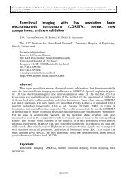
FIGURE 5- 4 Identificatio n <strong>of</strong> newly postmitotic cell s<br />
sensitive to apoptosis induced by inhibition <strong>of</strong> protein<br />
syn<strong>the</strong>sis. Immunolabeling for BrdU in <strong>the</strong> prolifera -<br />
tive zone o f a retinal expiant (arrows ) after inhibitio n<br />
<strong>of</strong> protein syn<strong>the</strong>sis. Notice that <strong>the</strong> majorit y o f dying<br />
cells (arrowheads) , identifie d a s globula r apoptoti c<br />
bodies under differential interferenc e contrast, i s unlabeled<br />
wit h BrdU . Scal e ba r = 20 \im. (Source:<br />
Adapted from Rehe n e t al., 1999 )<br />
CELL DEATH 7 9<br />
syn<strong>the</strong>sis i n th e developin g nervou s system <strong>and</strong> tar -<br />
geted deletio n o f <strong>the</strong> anti-apoptoti c gen e Bcl-x L suggest<br />
that during migration <strong>and</strong> initial differentiation,<br />
<strong>the</strong> PC D machiner y i s controlle d b y suppresso r<br />
proteins.<br />
PROLIFERATIVE CELL DEATH, DNA<br />
BREAKS AND END JOININ G<br />
Thymic PCD occur s during <strong>the</strong> selection o f functionally<br />
an d molecularl y distinc t thymocyte s (Sur h an d<br />
Sprent, 1994), based on somatic DNA rearrangements<br />
encoding T cell receptor s <strong>and</strong> immunoglobulins in T<br />
<strong>and</strong> B lymphocytes, respectively. This process, known<br />
as V(D)J recombination (Weaver <strong>and</strong> Alt, 1997) , i s responsible<br />
for <strong>the</strong> unparallele d degree <strong>of</strong> immunologicai<br />
diversit y tha t i s fundamenta l t o th e immun e<br />
response. A s describe d above , a paralle l exist s be -<br />
tween PCD i n <strong>the</strong> thymus <strong>and</strong> <strong>the</strong> feta l brain, where<br />
cell death takes place in conjunction with cell prolif -<br />
eration (Shortman et al, 1990 ; Chun, 2001).<br />
ISEL+ result s suggest that a form o f cell selectio n<br />
occurs i n th e neuroproliferativ e regions o f <strong>the</strong> brai n<br />
<strong>and</strong> that <strong>the</strong> occurrence <strong>of</strong> PCD i s associated with <strong>the</strong><br />
maturation proces s (Blaschk e e t al. , 1996 , 1998) . I n<br />
<strong>the</strong> feta l brain , <strong>the</strong> pea k <strong>of</strong> cell deat h coincide s wit h<br />
<strong>the</strong> perio d whe n th e first neurons destine d t o com -<br />
prise th e matur e corte x ar e bein g generate d (Cavi -<br />
ness, 1982) . Thi s spik e i n PC D migh t allo w th e<br />
selection o f <strong>the</strong> first cortical neuron s havin g desired<br />
phenotypes which woul d serv e as a template fo r th e<br />
selection o f later-generate d cells . Thi s hypo<strong>the</strong>tica l<br />
scenario leave s open th e questio n o f how cells migh t<br />
be mechanistically selected .<br />
Nearly a decade befor e geneti c recombinatio n was<br />
formally demonstrated in lymphocytes, Dreyer <strong>and</strong> colleagues<br />
(1967 ) postulate d tha t recombine d gene s en -<br />
coding cell-surface proteins might play a part in guiding<br />
<strong>the</strong> correct re-innervation <strong>of</strong> <strong>the</strong> goldfish tectum by retinal<br />
axons . These recombine d protein s would act a s a<br />
kind o f molecular cod e t o ensure that th e topographi -<br />
cally correc t retinotecta l mappin g i s achieved. Sinc e<br />
this idea was put forward, however, no gene undergoin g<br />
<strong>the</strong> requisit e recombination i n <strong>the</strong> nervou s system has<br />
been found . Thi s i s perhap s no t surprising , a s th e<br />
reagents that were instrumental in <strong>the</strong> discovery <strong>of</strong> lymphocyte<br />
V(D)J recombination—DNA sequences fro m<br />
a known c<strong>and</strong>idate locus, <strong>and</strong> clonal cell lines in which<br />
<strong>the</strong> rearrangement even t takes place—do not currently<br />
exist for <strong>the</strong> nervous system.