- Page 7 and 8:
Cambridge Botanical Handbooks Edite
- Page 9 and 10:
LICHENS BY ANNIE LORRAIN SMITH, F.L
- Page 11:
PREFACE THE publication of this vol
- Page 14 and 15:
viii CONTENTS II. LICHEN HYPHAE PAG
- Page 16 and 17:
x CONTENTS 2. RADIATE OR SECONDARY
- Page 18 and 19:
xii CONTENTS 2. PYRENOLICHENS a. De
- Page 20 and 21:
xiv CONTENTS C. SUPPLY OF INORGANIC
- Page 22 and 23:
xvi CONTENTS PAGE D. EVOLUTION OF A
- Page 24 and 25:
CONTENTS B. LICHENS AS MEDICINE ...
- Page 26 and 27:
xx GLOSSARY Discoid, disc-like, an
- Page 28 and 29:
xxii GLOSSARY Rhagadiose, deeply ch
- Page 30 and 31:
xxiv INTRODUCTION In the absence of
- Page 32 and 33:
xxvi INTRODUCTION the cortex have b
- Page 34 and 35:
xxviii INTRODUCTION species is a mo
- Page 36 and 37:
2 HISTORY OF LICHENOLOGY A seventh
- Page 38 and 39:
4 HISTORY OF LICHENOLOGY the specie
- Page 40 and 41:
6 HISTORY OF LICHENOLOGY plants are
- Page 42 and 43:
8 HISTORY OF LICHENOLOGY 5. "Coriac
- Page 44 and 45:
I0 HISTORY OF LICHENOLOGY Extensive
- Page 46 and 47:
12 HISTORY OF LICHENOLOGY Among con
- Page 48 and 49:
I4 HISTORY OF LICHENOLOGY he howeve
- Page 50 and 51:
16 HISTORY OF LICHENOLOGY departure
- Page 52 and 53:
I8 HISTORY OF LICHENOLOGY H. PERIOD
- Page 54 and 55:
CHAPTER II CONSTITUENTS OF THE LICH
- Page 56 and 57:
22 CONSTITUENTS OF THE LICHEN THALL
- Page 58 and 59:
24 CONSTITUENTS OF THE LICHEN THALL
- Page 60 and 61:
26 CONSTITUENTS OF THE LICHEN THALL
- Page 62 and 63:
CONSTITUENTS OF THE LICHEN THALLUS
- Page 64 and 65:
3o CONSTITUENTS OF THE LICHEN THALL
- Page 66 and 67:
3 2 CONSTITUENTS OF THE LICHEN THAL
- Page 68 and 69:
34 CONSTITUENTS OF THE LICHEN THALL
- Page 70 and 71:
36 CONSTITUENTS OF THE LICHEN THALL
- Page 72 and 73:
38 CONSTITUENTS OF THE LICHEN THALL
- Page 74 and 75:
40 CONSTITUENTS OF THE LICHEN THALL
- Page 76 and 77:
42 CONSTITUENTS OF THE LICHEN THALL
- Page 78 and 79:
44 CONSTITUENTS OF THE LICHEN THALL
- Page 80 and 81:
46 CONSTITUENTS OF THE LICHEN THALL
- Page 82 and 83:
48 CONSTITUENTS OF THE LICHEN THALL
- Page 84 and 85:
50 CONSTITUENTS OF THE LICHEN THALL
- Page 86 and 87:
CONSTITUENTS OF THE LICHEN THALLUS
- Page 88 and 89:
54 CONSTITUENTS OF THE LICHEN THALL
- Page 90 and 91:
CONSTITUENTS OF THE LICHEN THALLUS
- Page 92 and 93:
58 CONSTITUENTS OF THE LICHEN THALL
- Page 94 and 95:
6o CONSTITUENTS OF THE LICHEN THALL
- Page 96 and 97:
62 CONSTITUENTS OF THE LICHEN THALL
- Page 98 and 99:
64 CONSTITUENTS OF THE LICHEN THALL
- Page 100 and 101:
66 CONSTITUENTS OF THE LICHEN THALL
- Page 102 and 103:
68 MORPHOLOGY or chondroid strands
- Page 104 and 105:
;o MORPHOLOGY II. STRATOSE THALLUS
- Page 106 and 107:
72 MORPHOLOGY bb. Formation of crus
- Page 108 and 109:
74 MORPHOLOGY marked by subsequent
- Page 110 and 111:
;6 MORPHOLOGY c. CHEMICAL NATURE OF
- Page 112 and 113:
78 MORPHOLOGY b. HYPOPHLOEODAL LICH
- Page 114 and 115:
8o MORPHOLOGY species of Dermatocar
- Page 116 and 117:
82 3. MORPHOLOGY FOLIOSE LICHENS A.
- Page 118 and 119:
84 MORPHOLOGY in the molecular cons
- Page 120 and 121:
86 MORPHOLOGY become almost or enti
- Page 122 and 123:
88 MORPHOLOGY fore, in the most fav
- Page 124 and 125:
MORPHOLOGY b. LOWER CORTEX. In some
- Page 126 and 127:
9 2 MORPHOLOGY retain the colour of
- Page 128 and 129:
94 MORPHOLOGY or less characteristi
- Page 130 and 131:
96 MORPHOLOGY a. BY DEVELOPMENT OF
- Page 132 and 133:
9 8 MORPHOLOGY III. RADIATE THALLUS
- Page 134 and 135: 100 MORPHOLOGY In the two former th
- Page 136 and 137: 102 MORPHOLOGY thallus. It is also
- Page 138 and 139: 104 MORPHOLOGY sometimes as a conti
- Page 140: io6 MORPHOLOGY fastigiate cortex as
- Page 143 and 144: RADIATE THALLUS 109 afforded by the
- Page 145 and 146: RADIATE THALLUS in The sheath of R.
- Page 147 and 148: STRATOSE-RADIATE THALLUS 113 betwee
- Page 149 and 150: STRATOSE-RADIATE THALLUS thus disti
- Page 151 and 152: STRATOSE-RADIATE THALLUS 117 areas
- Page 153 and 154: STRATOSE-RADIATE THALLUS 119 follow
- Page 155 and 156: STRATOSE-RADIATE THALLUS 121 a cont
- Page 157 and 158: STRATOSE-RADIATE THALLUS 123 consid
- Page 159 and 160: STRATOSE-RADIATE THALLUS 125 the pr
- Page 161 and 162: STRUCTURES PECULIAR TO LICHENS 127
- Page 163 and 164: STRUCTURES PECULIAR TO LICHENS 129
- Page 165 and 166: STRUCTURES PECULIAR TO LICHENS 131
- Page 167 and 168: STRUCTURES PECULIAR TO LICHENS 133
- Page 169 and 170: STRUCTURES PECULIAR TO LICHENS 135
- Page 171 and 172: STRUCTURES PECULIAR TO LICHENS 137
- Page 173 and 174: STRUCTURES PECULIAR TO LICHENS 139
- Page 175 and 176: STRUCTURES PECULIAR TO LICHENS 141
- Page 177 and 178: STRUCTURES PECULIAR TO LICHENS of s
- Page 179 and 180: STRUCTURES PECULIAR TO LICHENS 145
- Page 181 and 182: STRUCTURES PECULIAR TO LICHENS 147
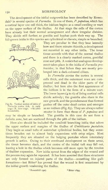
- Page 183: STRUCTURES PECULIAR TO LICHENS 149
- Page 187 and 188: HYMENOLICHENS 153 recognized its af
- Page 189 and 190: CHAPTER IV REPRODUCTION I. REPRODUC
- Page 191 and 192: REPRODUCTIVE ORGANS 157 size, rarel
- Page 193 and 194: REPRODUCTIVE ORGANS 159 is partiall
- Page 195 and 196: REPRODUCTION IN DISCOLICHENS the vi
- Page 197 and 198: REPRODUCTION IN DISCOLICHENS 163 or
- Page 199 and 200: REPRODUCTION IN DISCOLICHENS 165 fi
- Page 201 and 202: REPRODUCTION IN DISCOLICHENS 167 (2
- Page 203 and 204: REPRODUCTION IN DISCOLICHENS 169 ru
- Page 205 and 206: REPRODUCTION IN DISCOLICHENS 171 ti
- Page 207 and 208: REPRODUCTION IN DISCOLICHENS 173 br
- Page 209 and 210: APOGAMOUS REPRODUCTION 175 also by
- Page 211 and 212: DISCUSSION OF LICHEN REPRODUCTION 1
- Page 213 and 214: DISCUSSION OF LICHEN REPRODUCTION 1
- Page 215 and 216: DISCUSSION OF LICHEN REPRODUCTION 1
- Page 217 and 218: DEVELOPMENT OF APOTHECIA 183 wall o
- Page 219 and 220: LICHEN ASCI AND SPORES 185 b. DEVEL
- Page 221 and 222: LICHEN ASCI AND SPORES 187 than in
- Page 223 and 224: LICHEN ASCI AND SPORES 189 The deve
- Page 225 and 226: LICHEN ASCI AND SPORES 191 usually
- Page 227 and 228: SPERMOGONIA 193 B. SPERMOGONIA AS M
- Page 229 and 230: SPERMOGONIA 195 better lighted port
- Page 231 and 232: SPERMOGONIA 197 advanced stage the
- Page 233 and 234: SPERMOGONIA 199 "pycnidial" non-sex
- Page 235 and 236:
SPERMOGONIA 201 paraphyses, and als
- Page 237 and 238:
SPERMOGONIA 203 successful, was ver
- Page 239 and 240:
SPERMOGONIA 205 He also regards as
- Page 241 and 242:
SPERMOGONIA 207 Lichen " spermatia
- Page 243 and 244:
CHAPTER V PHYSIOLOGY I. CELLS AND C
- Page 245 and 246:
CELLS AND CELL PRODUCTS 211 colouri
- Page 247 and 248:
CELLS AND CELL PRODUCTS 213 B. CONT
- Page 249 and 250:
CELLS AND CELL PRODUCTS 215 pustula
- Page 251 and 252:
CELLS AND CELL PRODUCTS 217 slices
- Page 253 and 254:
CELLS AND CELL PRODUCTS 219 pected
- Page 255 and 256:
CELLS AND CELL PRODUCTS 221 an excr
- Page 257 and 258:
CELLS AND CELL PRODUCTS 223 not abo
- Page 259 and 260:
CELLS AND CELL PRODUCTS 225 the tha
- Page 261 and 262:
CELLS AND CELL PRODUCTS 227 lecanor
- Page 263 and 264:
CELLS AND CELL PRODUCTS 229 As an i
- Page 265 and 266:
GENERAL NUTRITION 231 c. FOLIOSE LI
- Page 267 and 268:
GENERAL NUTRITION 233 from atmosphe
- Page 269 and 270:
GENERAL NUTRITION 235 part of an ol
- Page 271 and 272:
GENERAL NUTRITION 237 Lecanora thal
- Page 273 and 274:
ASSIMILATION AND RESPIRATION 239 Ju
- Page 275 and 276:
ILLUMINATION OF LICHENS 241 Wiesner
- Page 277 and 278:
ILLUMINATION OF LICHENS 243 shade i
- Page 279 and 280:
ILLUMINATION OF LICHENS 245 inserti
- Page 281 and 282:
COLOUR OF LICHENS 247 In many cases
- Page 283 and 284:
COLOUR OF LICHENS 249 apothecia of
- Page 285 and 286:
251
- Page 287 and 288:
GROWTH AND DURATION 253 and suggest
- Page 289 and 290:
GROWTH AND DURATION 255 in the same
- Page 291 and 292:
DISPERSAL AND INCREASE 257 Crustace
- Page 293 and 294:
ERRATIC LICHENS 259 bind the mass t
- Page 295 and 296:
PARASITISM 261 other lichens. The c
- Page 297 and 298:
PARASITISM 263 there on the thallus
- Page 299 and 300:
PARASITISM 265 and found that they
- Page 301 and 302:
PARASITISM 267 observed. The mature
- Page 303 and 304:
DISEASES OF LICHENS 269 the upper c
- Page 305 and 306:
GALL-FORMATION 271 irritation excit
- Page 307 and 308:
ORIGIN OF LICHENS 273 B. ALGAL ANCE
- Page 309 and 310:
REPRODUCTIVE ORGANS 275 B. RELATION
- Page 311 and 312:
REPRODUCTIVE ORGANS 277 they are mo
- Page 313 and 314:
REPRODUCTIVE ORGANS 279 as " hypoph
- Page 315 and 316:
REPRODUCTIVE ORGANS 281 lichen genu
- Page 317 and 318:
THE THALLUS 283 might become fertil
- Page 319 and 320:
THE THALLUS 285 there is but little
- Page 321 and 322:
THE THALLUS 287 Peltigera and Nephr
- Page 323 and 324:
THE THALLUS 289 genus Pyrgillus has
- Page 325 and 326:
THE THALLUS 291 aa. COENOGONIACEAE.
- Page 327 and 328:
THE THALLUS 293 are colourless and
- Page 329 and 330:
THE THALLUS 295 of the scyphus and
- Page 331 and 332:
THE THALLUS 297 His third group inc
- Page 333 and 334:
THE THALLUS 299 are composed of com
- Page 335 and 336:
THE THALLUS 301 species and forms a
- Page 337 and 338:
THE THALLUS 303 SCHEME OF SUGGESTED
- Page 339 and 340:
FAMILIES AND GENERA 305 The apothec
- Page 341 and 342:
FAMILIES AND GENERA 307 based on im
- Page 343 and 344:
FAMILIES AND GENERA 309 SERifcs i.
- Page 345 and 346:
FAMILIES AND GENERA 311 b. Not gela
- Page 347 and 348:
FAMILIES AND GENERA 313 "pycnidia"
- Page 349 and 350:
FAMILIES AND GENERA 315 The gonidia
- Page 351 and 352:
FAMILIES AND GENERA 317 superficial
- Page 353 and 354:
FAMILIES AND GENERA 319 XIV. PYRENW
- Page 355 and 356:
FAMILIES AND GENERA 321 graplia} re
- Page 357 and 358:
FAMILIES AND GENERA 323 Thallus wit
- Page 359 and 360:
FAMILIES AND GENERA 325 XXIII. LECA
- Page 361 and 362:
FAMILIES AND GENERA 327 XXVIII. ECT
- Page 363 and 364:
Thallus crustaceous non-corticate.
- Page 365 and 366:
FAMILIES AND GENERA 331 which grows
- Page 367 and 368:
Thallus with Gloeocapsa gonidia. Th
- Page 369 and 370:
FAMILIES AND GENERA 335 XL. HEPPIAC
- Page 371 and 372:
FAMILIES AND GENERA 337 margin of t
- Page 373 and 374:
Thallus non-corticate below. FAMILI
- Page 375 and 376:
FAMILIES AND GENERA 341 XLIX. TELQS
- Page 377 and 378:
NUMBER OF LICHENS 343 or described
- Page 379 and 380:
DISTRIBUTION 345 but, as yet, has b
- Page 381 and 382:
DISTRIBUTION 347 as Cladoniae, Leca
- Page 383 and 384:
DISTRIBUTION 349 its habitat as "th
- Page 385 and 386:
DISTRIBUTION 351 lichens of Austral
- Page 387 and 388:
DISTRIBUTION 353 epiphyllous lichen
- Page 389 and 390:
FOSSIL LICHENS 355 which recalls so
- Page 391 and 392:
GENERAL INTRODUCTION 357 Ecological
- Page 393 and 394:
EXTERNAL INFLUENCES 359 observed th
- Page 395 and 396:
EXTERNAL INFLUENCES 361 South Lanca
- Page 397 and 398:
LICHEN COMMUNITIES 363 I. ARBOREAL
- Page 399 and 400:
LICHEN COMMUNITIES 365 whether of y
- Page 401 and 402:
LICHEN COMMUNITIES 367 gained a foo
- Page 403 and 404:
LICHEN COMMUNITIES 369 On bare heat
- Page 405 and 406:
LICHEN COMMUNITIES 371 As already d
- Page 407 and 408:
LICHEN COMMUNITIES 373 Most calcico
- Page 409 and 410:
LICHEN COMMUNITIES 375 It will only
- Page 411 and 412:
LICHEN COMMUNITIES 377 The followin
- Page 413 and 414:
indifferently on any LICHEN COMMUNI
- Page 415 and 416:
LICHEN COMMUNITIES 123. Ramalina si
- Page 417 and 418:
LICHEN COMMUNITIES 383 width all ro
- Page 419 and 420:
LICHEN COMMUNITIES: 385 grass that
- Page 421 and 422:
LICHEN COMMUNITIES 387 above the ti
- Page 423 and 424:
LICHEN COMMUNITIES - C. crispa are
- Page 425 and 426:
LICHEN COMMUNITIES 391 country is D
- Page 427 and 428:
LICHENS AS PIONEERS 393 action of h
- Page 429 and 430:
CHAPTER X ECONOMIC AND TECHNICAL A.
- Page 431 and 432:
are, however, not equally palatable
- Page 433 and 434:
LICHENS AS FOOD 399 A minute organi
- Page 435 and 436:
LICHENS AS FOOD 401 The true reinde
- Page 437 and 438:
LICHENS AS FOOD 403 Iso-lichenin is
- Page 439 and 440:
LICHENS AS FOOD 405 20 cm. with irr
- Page 441 and 442:
LICHENS AS MEDICINE 407 The doctrin
- Page 443 and 444:
LICHENS AS MEDICINE 409 directions
- Page 445 and 446:
LICHENS IN INDUSTRY 411 D. LICHENS
- Page 447 and 448:
LICHENS AS DYE-PLANTS 413 British s
- Page 449 and 450:
LICHENS AS DYE-PLANTS 415 of rock-
- Page 451 and 452:
LICHENS AS DYE-PLANTS 417 boiling.
- Page 453 and 454:
LICHENS IN PERFUMERY 419 b. LICHENS
- Page 455 and 456:
APPENDIX POSTSCRIPT TO CHAPTER VII
- Page 457 and 458:
BIBLIOGRAPHY OF BOOKS AND PAPERS CI
- Page 459 and 460:
BIBLIOGRAPHY 425 Beyerinck, M. W. D
- Page 461 and 462:
BIBLIOGRAPHY 427 Crombie, J. M. On
- Page 463 and 464:
BIBLIOGRAPHY 429 Etard and Bouilhac
- Page 465 and 466:
BIBLIOGRAPHY 431 Gibelli, Giuseppe.
- Page 467 and 468:
BIBLIOGRAPHY 433 Hudson, W. Flora A
- Page 469 and 470:
BIBLIOGRAPHY 435 Krempelhuber, Augu
- Page 471 and 472:
BIBLIOGRAPHY 437 Malinowski, E. Sur
- Page 473 and 474:
BIBLIOGRAPHY 439 Nylander, W. Consp
- Page 475 and 476:
BIBLIOGRAPHY 441 Rosendahl, F. Verg
- Page 477 and 478:
BIBLIOGRAPHY 443 Steiner, J. Beitra
- Page 479 and 480:
BIBLIOGRAPHY 445 Watson, Sir W. An
- Page 481 and 482:
BIBLIOGRAPHY 447 Zukal, H. Das Zusa
- Page 483 and 484:
Bacotia septum, 399 Baeotnyces Pers
- Page 485 and 486:
INDEX Cladonia sqiiamosa Hoffm., 11
- Page 487 and 488:
Forssellia A. Zahlbr., 284, 333, 37
- Page 489 and 490:
Lecanora dtrina Ach., see Placodium
- Page 491 and 492:
Mereschkovsky, 258 Merrett, 3 Metzg
- Page 493 and 494:
Pentagenella Darbish., 83, 324 Perf
- Page 495 and 496:
Quercus alba, 359 Q. chrysolepis, 3
- Page 497 and 498:
Stigonema Ag., 23, 16, 54 (Fig. 20)
- Page 502 and 503:
UNIVERSITY OF CALIFORNIA LIBRARY Lo


