

Strabismus - Fundamentals of Clinical Ophthalmology.pdf
Strabismus - Fundamentals of Clinical Ophthalmology.pdf
Strabismus - Fundamentals of Clinical Ophthalmology.pdf

You also want an ePaper? Increase the reach of your titles
YUMPU automatically turns print PDFs into web optimized ePapers that Google loves.
2 A simple reflex model <strong>of</strong><br />
normal binocular vision<br />
Introduction<br />
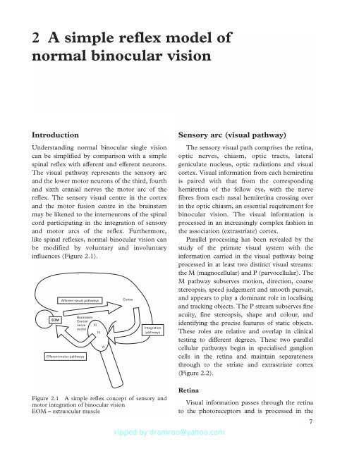
Understanding normal binocular single vision<br />
can be simplified by comparison with a simple<br />
spinal reflex with afferent and efferent neurons.<br />
The visual pathway represents the sensory arc<br />
and the lower motor neurons <strong>of</strong> the third, fourth<br />
and sixth cranial nerves the motor arc <strong>of</strong> the<br />
reflex. The sensory visual centre in the cortex<br />
and the motor fusion centre in the brainstem<br />
may be likened to the interneurons <strong>of</strong> the spinal<br />
cord participating in the integration <strong>of</strong> sensory<br />
and motor arcs <strong>of</strong> the reflex. Furthermore,<br />
like spinal reflexes, normal binocular vision can<br />
be modified by voluntary and involuntary<br />
influences (Figure 2.1).<br />
EOM<br />
Afferent visual pathways<br />
Brainstem<br />
Cranial<br />
nerve<br />
nuclei<br />
Efferent motor pathways<br />
III<br />
IV<br />
Cortex<br />
Integration<br />
pathways<br />
Figure 2.1 A simple reflex concept <strong>of</strong> sensory and<br />
motor integration <strong>of</strong> binocular vision<br />
EOM = extraocular muscle<br />
VI<br />
Sensory arc (visual pathway)<br />
The sensory visual path comprises the retina,<br />
optic nerves, chiasm, optic tracts, lateral<br />
geniculate nucleus, optic radiations and visual<br />
cortex. Visual information from each hemiretina<br />
is paired with that from the corresponding<br />
hemiretina <strong>of</strong> the fellow eye, with the nerve<br />
fibres from each nasal hemiretina crossing over<br />
in the optic chiasm, an essential requirement for<br />
binocular vision. The visual information is<br />
processed in an increasingly complex fashion in<br />
the association (extrastriate) cortex.<br />
Parallel processing has been revealed by the<br />
study <strong>of</strong> the primate visual system with the<br />
information carried in the visual pathway being<br />
processed in at least two distinct visual streams:<br />
the M (magnocellular) and P (parvocellular). The<br />
M pathway subserves motion, direction, coarse<br />
stereopsis, speed judgement and smooth pursuit,<br />
and appears to play a dominant role in localising<br />
and tracking objects. The P stream subserves fine<br />
acuity, fine stereopsis, shape and colour, and<br />
identifying the precise features <strong>of</strong> static objects.<br />
These roles are relative and overlap in clinical<br />
testing to different degrees. These two parallel<br />
cellular pathways begin in specialised ganglion<br />
cells in the retina and maintain separateness<br />
through to the striate and extrastriate cortex<br />
(Figure 2.2).<br />
Retina<br />
Visual information passes through the retina<br />
to the photoreceptors and is processed in the<br />
7


![SISTEM SENSORY [Compatibility Mode].pdf](https://img.yumpu.com/20667975/1/190x245/sistem-sensory-compatibility-modepdf.jpg?quality=85)


