

Strabismus - Fundamentals of Clinical Ophthalmology.pdf
Strabismus - Fundamentals of Clinical Ophthalmology.pdf
Strabismus - Fundamentals of Clinical Ophthalmology.pdf

Create successful ePaper yourself
Turn your PDF publications into a flip-book with our unique Google optimized e-Paper software.
STRABISMUS<br />
Table 2.2<br />
Types <strong>of</strong> eye movements and functions<br />
Type <strong>of</strong> eye movement<br />
Vestibular<br />
Optokinetic<br />
Saccade<br />
Smooth pursuit<br />
Vergence<br />
Visual fixation<br />
Function<br />
Maintains image on retina during<br />
head rotation (brief)<br />
Maintains image on retina during<br />
head rotation (sustained)<br />
Captures fresh objects <strong>of</strong> interest<br />
on fovea – refixation<br />
Maintains image <strong>of</strong> moving target<br />
on fovea – sustains fixation<br />
Maintains image <strong>of</strong> a single<br />
object through disjunctive<br />
movement<br />
Maintains clear visual image on<br />
the fovea with head stationary<br />
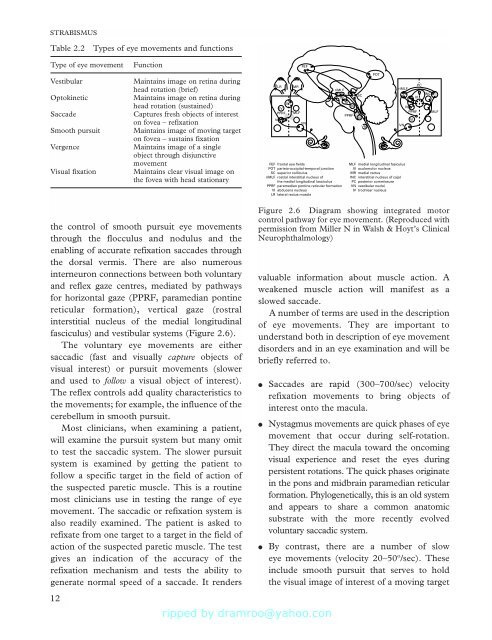
FEF<br />
POT<br />
LR MR<br />
PC<br />
riMLF<br />
riMLF<br />
SC<br />
INC<br />
INC<br />
III<br />
III<br />
III<br />
IV<br />
IV<br />
IV<br />
PPRF MLF<br />
PPRF MLF<br />
PPRF<br />
VI<br />
VI<br />
VI<br />
VN<br />
VN<br />
VN<br />
FEF frontal eye fields<br />
MLF medial longitudinal fasiculus<br />
POT parieto-occipital-temporal junction<br />
III oculomolor nucleus<br />
SC superior colliculus<br />
MR medial rectus<br />
riMLF rostral interstitial nucleus <strong>of</strong><br />
INC interstitial nucleus <strong>of</strong> cajal<br />
the medial longitudinal lasciculus<br />
PC posterior commissure<br />
PPRF paramedian pontine reticular formation VN vestibular nuclei<br />
VI abducens nucleus<br />
IV trochlear nucleus<br />
LR lateral rectus muscle<br />
the control <strong>of</strong> smooth pursuit eye movements<br />
through the flocculus and nodulus and the<br />
enabling <strong>of</strong> accurate refixation saccades through<br />
the dorsal vermis. There are also numerous<br />
interneuron connections between both voluntary<br />
and reflex gaze centres, mediated by pathways<br />
for horizontal gaze (PPRF, paramedian pontine<br />
reticular formation), vertical gaze (rostral<br />
interstitial nucleus <strong>of</strong> the medial longitudinal<br />
fasciculus) and vestibular systems (Figure 2.6).<br />
The voluntary eye movements are either<br />
saccadic (fast and visually capture objects <strong>of</strong><br />
visual interest) or pursuit movements (slower<br />
and used to follow a visual object <strong>of</strong> interest).<br />
The reflex controls add quality characteristics to<br />
the movements; for example, the influence <strong>of</strong> the<br />
cerebellum in smooth pursuit.<br />
Most clinicians, when examining a patient,<br />
will examine the pursuit system but many omit<br />
to test the saccadic system. The slower pursuit<br />
system is examined by getting the patient to<br />
follow a specific target in the field <strong>of</strong> action <strong>of</strong><br />
the suspected paretic muscle. This is a routine<br />
most clinicians use in testing the range <strong>of</strong> eye<br />
movement. The saccadic or refixation system is<br />
also readily examined. The patient is asked to<br />
refixate from one target to a target in the field <strong>of</strong><br />
action <strong>of</strong> the suspected paretic muscle. The test<br />
gives an indication <strong>of</strong> the accuracy <strong>of</strong> the<br />
refixation mechanism and tests the ability to<br />
generate normal speed <strong>of</strong> a saccade. It renders<br />
12<br />
Figure 2.6 Diagram showing integrated motor<br />
control pathway for eye movement. (Reproduced with<br />
permission from Miller N in Walsh & Hoyt’s <strong>Clinical</strong><br />
Neurophthalmology)<br />
valuable information about muscle action. A<br />
weakened muscle action will manifest as a<br />
slowed saccade.<br />
A number <strong>of</strong> terms are used in the description<br />
<strong>of</strong> eye movements. They are important to<br />
understand both in description <strong>of</strong> eye movement<br />
disorders and in an eye examination and will be<br />
briefly referred to.<br />
●<br />
●<br />
●<br />
Saccades are rapid (300–700/sec) velocity<br />
refixation movements to bring objects <strong>of</strong><br />
interest onto the macula.<br />
Nystagmus movements are quick phases <strong>of</strong> eye<br />
movement that occur during self-rotation.<br />
They direct the macula toward the oncoming<br />
visual experience and reset the eyes during<br />
persistent rotations. The quick phases originate<br />
in the pons and midbrain paramedian reticular<br />
formation. Phylogenetically, this is an old system<br />
and appears to share a common anatomic<br />
substrate with the more recently evolved<br />
voluntary saccadic system.<br />
By contrast, there are a number <strong>of</strong> slow<br />
eye movements (velocity 20–50º/sec). These<br />
include smooth pursuit that serves to hold<br />
the visual image <strong>of</strong> interest <strong>of</strong> a moving target


![SISTEM SENSORY [Compatibility Mode].pdf](https://img.yumpu.com/20667975/1/190x245/sistem-sensory-compatibility-modepdf.jpg?quality=85)


