

Strabismus - Fundamentals of Clinical Ophthalmology.pdf
Strabismus - Fundamentals of Clinical Ophthalmology.pdf
Strabismus - Fundamentals of Clinical Ophthalmology.pdf

You also want an ePaper? Increase the reach of your titles
YUMPU automatically turns print PDFs into web optimized ePapers that Google loves.
Transient skew deviation<br />
CHILDHOOD ONSET OF STRABISMUS<br />
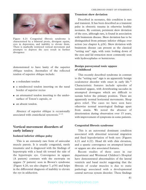
Figure 4.21 Congenital fibrosis syndrome is<br />
characterised by a bilateral ptosis, divergent squint,<br />
eyes in depression, and inability to elevate them.<br />
There is markedly restricted vertical movement and<br />
attempts to depress the eyes result in further<br />
divergence<br />
Described in neonates, this condition is rare<br />
and transient. It has been described as a transient<br />
palsy in obstetric trauma in otherwise healthy<br />
neonates. By contrast, persistent skew deviation<br />
<strong>of</strong> the eyes, although rare, is found in association<br />
with brainstem disease. Skew deviation has to be<br />
distinguished from primary inferior oblique over<br />
action (see chapter 4, p26). Skew deviation with<br />
brainstem disease can present as the classical<br />
“setting sun” sign, with tonic looking down <strong>of</strong><br />
the eyes and lid retraction most commonly seen<br />
with hydrocephalus or kernicterus.<br />
demonstrated to have laxity <strong>of</strong> the superior<br />
oblique tendon. Anomalies <strong>of</strong> the reflected<br />
tendon <strong>of</strong> superior oblique include:<br />
●<br />
●<br />
●<br />
●<br />
a redundant tendon<br />
a misdirected tendon inserting on the nasal<br />
border <strong>of</strong> superior rectus<br />
an attenuated tendon inserting to the undersurface<br />
<strong>of</strong> Tenon’s capsule, or<br />
an absent tendon.<br />
Absence <strong>of</strong> superior oblique is occasionally<br />
associated with crani<strong>of</strong>acial synostosis. 23–25<br />
Benign paroxysmal tonic upgaze<br />
<strong>of</strong> childhood<br />
This recently described syndrome in contrast<br />
to the “setting sun” sign is an apparently benign<br />
oculomotor disorder with onset in early life. 26<br />
Characteristic features include episodes <strong>of</strong><br />
sustained upgaze, with downbeating saccades in<br />
attempted downgaze which are difficult to<br />
sustain below the primary position. There are<br />
apparently normal horizontal movements. Sleep<br />
gives relief. The cases we have seen have<br />
otherwise normal neurological findings apart<br />
from ataxia. We noted the absence <strong>of</strong><br />
deterioration during observation over 15 years,<br />
with improvement <strong>of</strong> symptoms in some patients.<br />
Vertical movement disorders <strong>of</strong><br />
early infancy<br />
Isolated inferior oblique palsy<br />
This is an extremely rare form <strong>of</strong> uniocular<br />
muscle paresis. It is usually congenital, rarely<br />
traumatic and is diagnosed with the findings <strong>of</strong><br />
hypertropia with a head tilt toward the side <strong>of</strong><br />
the hypertropic eye. Convergence in upgaze<br />
(A pattern) contrasts with the exotropia on<br />
upgaze (V pattern) seen in Brown’s syndrome<br />
(Figure 4.20, see also chapter 7, p78) and helps<br />
in the differential diagnosis <strong>of</strong> inability to elevate<br />
the eye in adduction.<br />
Congenital fibrosis syndrome<br />
This is an autosomal dominant condition<br />
associated with abnormal neuronal migration<br />
and fixed hypodeviation <strong>of</strong> both eyes (Figures<br />
4.21 and 4.22). Head tilt with chin elevation,<br />
and a spastic convergence on attempted lateral<br />
or upgaze are also associated features.<br />
Recent studies <strong>of</strong> these cases in our<br />
department and Westmead Children’s Hospital<br />
have demonstrated abnormalities <strong>of</strong> the lateral<br />
ventricle and basal nuclei suggesting that the<br />
fibrosis <strong>of</strong> ocular muscles is due to static<br />
pathology associated with a developmental<br />
central nervous system disorder. These findings<br />
41


![SISTEM SENSORY [Compatibility Mode].pdf](https://img.yumpu.com/20667975/1/190x245/sistem-sensory-compatibility-modepdf.jpg?quality=85)


