Die Embryonalentwicklung der Paradiesschnecke ... - TOBIAS-lib
Die Embryonalentwicklung der Paradiesschnecke ... - TOBIAS-lib
Die Embryonalentwicklung der Paradiesschnecke ... - TOBIAS-lib
You also want an ePaper? Increase the reach of your titles
YUMPU automatically turns print PDFs into web optimized ePapers that Google loves.
Kapitel 4<br />
Development of the nervous system in untorted and “normal” M.<br />
cornuarietis<br />
Examinations of embryos incubated with anti-FMRFamide and anti-tubulin<br />
antibodies showed little specific staining and the results are not shown. Thus,<br />
in the following, we exclusively focus on serotonin-like immunoreactivity in<br />
controls and Pt 2+ -exposed embryos. The application of an extended clearing<br />
using the ScaleB4-mixture proved to be essential for the visualization of the<br />
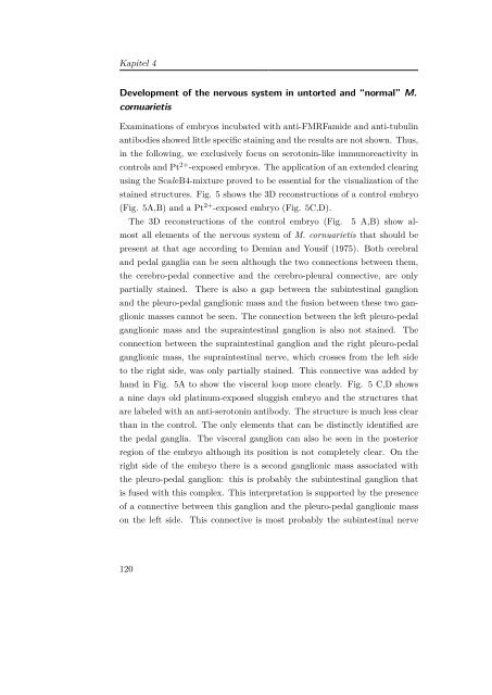
stained structures. Fig. 5 shows the 3D reconstructions of a control embryo<br />
(Fig. 5A,B) and a Pt 2+ -exposed embryo (Fig. 5C,D).<br />
The 3D reconstructions of the control embryo (Fig.<br />
5 A,B) show almost<br />
all elements of the nervous system of M. cornuarietis that should be<br />
present at that age according to Demian and Yousif (1975). Both cerebral<br />
and pedal ganglia can be seen although the two connections between them,<br />
the cerebro-pedal connective and the cerebro-pleural connective, are only<br />
partially stained.<br />
There is also a gap between the subintestinal ganglion<br />
and the pleuro-pedal ganglionic mass and the fusion between these two ganglionic<br />
masses cannot be seen. The connection between the left pleuro-pedal<br />
ganglionic mass and the supraintestinal ganglion is also not stained. The<br />
connection between the supraintestinal ganglion and the right pleuro-pedal<br />
ganglionic mass, the supraintestinal nerve, which crosses from the left side<br />
to the right side, was only partially stained. This connective was added by<br />
hand in Fig. 5A to show the visceral loop more clearly. Fig. 5 C,D shows<br />
a nine days old platinum-exposed sluggish embryo and the structures that<br />
are labeled with an anti-serotonin antibody. The structure is much less clear<br />
than in the control. The only elements that can be distinctly identified are<br />
the pedal ganglia. The visceral ganglion can also be seen in the posterior<br />
region of the embryo although its position is not completely clear. On the<br />
right side of the embryo there is a second ganglionic mass associated with<br />
the pleuro-pedal ganglion: this is probably the subintestinal ganglion that<br />
is fused with this complex. This interpretation is supported by the presence<br />
of a connective between this ganglion and the pleuro-pedal ganglionic mass<br />
on the left side. This connective is most probably the subintestinal nerve<br />
120


