

Die Embryonalentwicklung der Paradiesschnecke ... - TOBIAS-lib
Die Embryonalentwicklung der Paradiesschnecke ... - TOBIAS-lib
Die Embryonalentwicklung der Paradiesschnecke ... - TOBIAS-lib

You also want an ePaper? Increase the reach of your titles
YUMPU automatically turns print PDFs into web optimized ePapers that Google loves.
Kapitel 2<br />
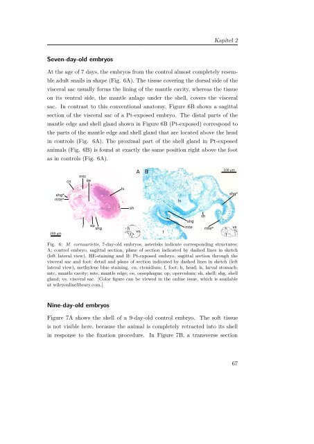
Seven-day-old embryos<br />
At the age of 7 days, the embryos from the control almost completely resemble<br />
adult snails in shape (Fig. 6A). The tissue covering the dorsal side of the<br />
visceral sac usually forms the lining of the mantle cavity, whereas the tissue<br />
on its ventral side, the mantle anlage un<strong>der</strong> the shell, covers the visceral<br />
sac. In contrast to this conventional anatomy, Figure 6B shows a sagittal<br />
section of the visceral sac of a Pt-exposed embryo. The distal parts of the<br />
mantle edge and shell gland shown in Figure 6B (Pt-exposed) correspond to<br />
the parts of the mantle edge and shell gland that are located above the head<br />
in controls (Fig. 6A). The proximal part of the shell gland in Pt-exposed<br />
animals (Fig. 6B) is found at exactly the same position right above the foot<br />
as in controls (Fig. 6A).<br />
Fig. 6: M. cornuarietis, 7-day-old embryos, asterisks indicate corresponding structures;<br />
A: control embryo, sagittal section, plane of section indicated by dashed lines in sketch<br />
(left lateral view), HE-staining and B: Pt-exposed embryo, sagittal section through the<br />
visceral sac and foot; detail and plane of section indicated by dashed lines in sketch (left<br />
lateral view), methylene blue staining. cn, ctenidium; f, foot; h, head; ls, larval stomach;<br />
mtc, mantle cavity; mte, mantle edge; oe, oesophagus; op, operculum; sh, shell; shg, shell<br />
gland; vs, visceral sac. [Color figure can be viewed in the online issue, which is available<br />
at wileyonline<strong>lib</strong>rary.com.]<br />
Nine-day-old embryos<br />
Figure 7A shows the shell of a 9-day-old control embryo. The soft tissue<br />
is not visible here, because the animal is completely retracted into its shell<br />
in response to the fixation procedure. In Figure 7B, a transverse section<br />
67


