
Create successful ePaper yourself
Turn your PDF publications into a flip-book with our unique Google optimized e-Paper software.
Organization of the Brain: Cell Types<br />
NEUROPHYSIOLOGY<br />
Multipolar (pyramidal)<br />
cell of cerebral<br />
motor cortex<br />
Astrocyte<br />
Motor<br />
endplate<br />
Multipolar somatic<br />
motor cell of nuclei<br />
of cranial nn.<br />
Multipolar cell<br />
of lower brain<br />
motor centers<br />
Oligodendrocyte<br />
Corticospinal<br />
(pyramidal) fiber<br />
Axodendritic ending<br />
Axosomatic ending<br />
Axoaxonic ending<br />
Multipolar somatic<br />
motor cell of<br />
anterior horn<br />
of spinal cord<br />
Collateral<br />
Striated<br />
(somatic)<br />
muscle<br />
Renshaw interneuron<br />
(feedback)<br />
Myelinated somatic motor<br />
fiber of spinal nerve<br />
Interneurons<br />
Blood vessel<br />
Interneuron<br />
Astrocyte<br />
Multipolar visceral<br />
motor (autonomic)<br />
cell of spinal cord<br />
Autonomic preganglionic<br />
(sympathetic or parasympathetic)<br />
nerve fiber<br />
Myelin sheath<br />
Autonomic postganglionic<br />
neuron of sympathetic or<br />
parasympathetic ganglion<br />
Satellite cells<br />
Unmyelinated nerve fiber<br />
Schwann cells<br />
Bipolar cell of cranial n.<br />
Unipolar cell of<br />
sensory ganglia of<br />
cranial nn.<br />
Satellite cells<br />
Schwann cell<br />
Free nerve endings<br />
(unmyelinated fibers)<br />
Encapsulated ending<br />
Specialized ending<br />
Muscle spindle<br />
Unipolar sensory cell<br />
of dorsal spinal<br />
root ganglion<br />
Satellite cells<br />
Myelinated afferent<br />
fiber of spinal nerve<br />
Myelin sheath<br />
Red: Motor neuron<br />
Blue: Sensory neuron<br />
Purple: Interneuron<br />
Gray: Glial and<br />
neurilemmal cells<br />
and myelin<br />
Note: Cerebellar cells<br />
not shown here<br />
Myelin sheath<br />
Schwann cells<br />
Motor endplate with<br />
Schwann cell cap<br />
Striated<br />
(voluntary)<br />
muscle<br />
Endings on<br />
cardiac muscle<br />
or nodal cells<br />
Beaded<br />
varicosities<br />
and endings on<br />
smooth muscle<br />
and gland cells<br />
Unmyelinated fibers<br />
Free nerve endings<br />
Encapsulated ending<br />
Muscle spindle<br />
©<br />
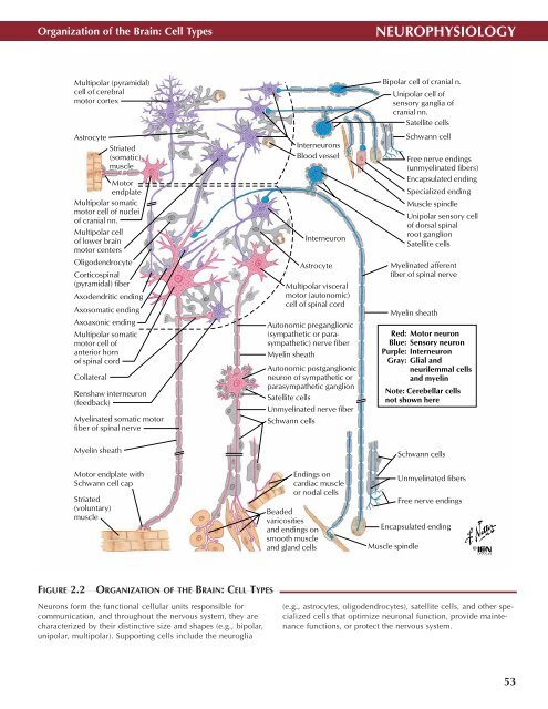
FIGURE 2.2<br />
ORGANIZATION OF THE BRAIN: CELL TYPES •<br />
<strong>Neuro</strong>ns form the functional cellular units responsible for<br />
communication, and throughout the nervous system, they are<br />
characterized by their distinctive size and shapes (e.g., bipolar,<br />
unipolar, multipolar). Supporting cells include the neuroglia<br />
(e.g., astrocytes, oligodendrocytes), satellite cells, and other specialized<br />
cells that optimize neuronal function, provide maintenance<br />
functions, or protect the nervous system.<br />
53